7OJN | pdb_00007ojn

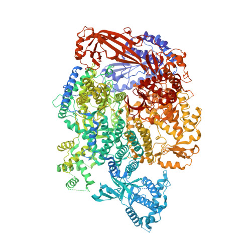
Lassa virus L protein in an elongation conformation [ELONGATION]


Experimental Data Snapshot

  • Method: ELECTRON MICROSCOPY
  • Resolution: 2.92 Å
  • Aggregation State: PARTICLE 
  • Reconstruction Method: SINGLE PARTICLE 

wwPDB Validation 3D Report Full Report

Validation slider image for 7OJN

This is version 1.3 of the entry. See complete history. 

Literature

Conformational changes in Lassa virus L protein associated with promoter binding and RNA synthesis activity.

Kouba, T., Vogel, D., Thorkelsson, S.R., Quemin, E.R.J., Williams, H.M., Milewski, M., Busch, C., Gunther, S., Grunewald, K., Rosenthal, M., Cusack, S.

(2021) Nat Commun 12: 7018-7018

  • DOI: https://doi.org/10.1038/s41467-021-27305-5
  • Primary Citation Related Structures: 
    7OCH, 7OE3, 7OE7, 7OEA, 7OEB, 7OJJ, 7OJK, 7OJL, 7OJN

  • PubMed Abstract: 

    Lassa virus is endemic in West Africa and can cause severe hemorrhagic fever. The viral L protein transcribes and replicates the RNA genome via its RNA-dependent RNA polymerase activity. Here, we present nine cryo-EM structures of the L protein in the apo-, promoter-bound pre-initiation and active RNA synthesis states. We characterize distinct binding pockets for the conserved 3' and 5' promoter RNAs and show how full-promoter binding induces a distinct pre-initiation conformation. In the apo- and early elongation states, the endonuclease is inhibited by two distinct L protein peptides, whereas in the pre-initiation state it is uninhibited. In the early elongation state, a template-product duplex is bound in the active site cavity together with an incoming non-hydrolysable nucleotide and the full C-terminal region of the L protein, including the putative cap-binding domain, is well-ordered. These data advance our mechanistic understanding of how this flexible and multifunctional molecular machine is activated.


  • Organizational Affiliation: 
    • European Molecular Biology Laboratory, Grenoble, France.

Macromolecule Content 

  • Total Structure Weight: 275.93 kDa 
  • Atom Count: 16,991 
  • Modeled Residue Count: 2,049 
  • Deposited Residue Count: 2,284 
  • Unique protein chains: 1
  • Unique nucleic acid chains: 3

Macromolecules


Find similar proteins by:|  3D Structure
Entity ID: 1
MoleculeChains  Sequence LengthOrganismDetailsImage
RNA-directed RNA polymerase LA [auth L]2,217Mammarenavirus lassaenseMutation(s): 0 
EC: 2.7.7.48 (PDB Primary Data), 3.1 (PDB Primary Data)
UniProt
Find proteins for A0A3S8NV63 (Mammarenavirus lassaense)
Explore A0A3S8NV63 
Go to UniProtKB:  A0A3S8NV63
Entity Groups
Sequence Clusters30% Identity50% Identity70% Identity90% Identity95% Identity100% Identity
UniProt GroupA0A3S8NV63
Sequence Annotations
Expand
Reference Sequence
Find similar nucleic acids by:  Sequence
Entity ID: 2
MoleculeChains LengthOrganismImage
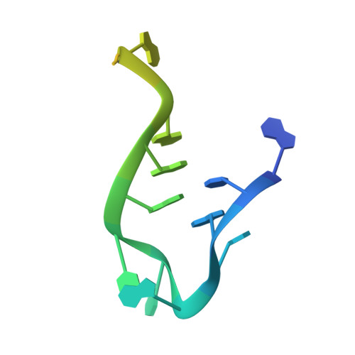
5' RNAB [auth D]20Mammarenavirus lassaense
Sequence Annotations
Expand
Reference Sequence
Find similar nucleic acids by:  Sequence
Entity ID: 3
MoleculeChains LengthOrganismImage
3' RNAC [auth R],
E
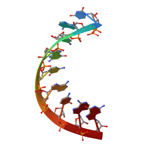
19Mammarenavirus lassaense
Sequence Annotations
Expand
Reference Sequence
Find similar nucleic acids by:  Sequence
Entity ID: 4
MoleculeChains LengthOrganismImage
product RNAD [auth M]9Mammarenavirus lassaense
Sequence Annotations
Expand
Reference Sequence

Small Molecules

Ligands 3 Unique
IDChains Name / Formula / InChI Key2D Diagram3D Interactions
2KH
(Subject of Investigation/LOI)

Query on 2KH


Download:Ideal Coordinates CCD File
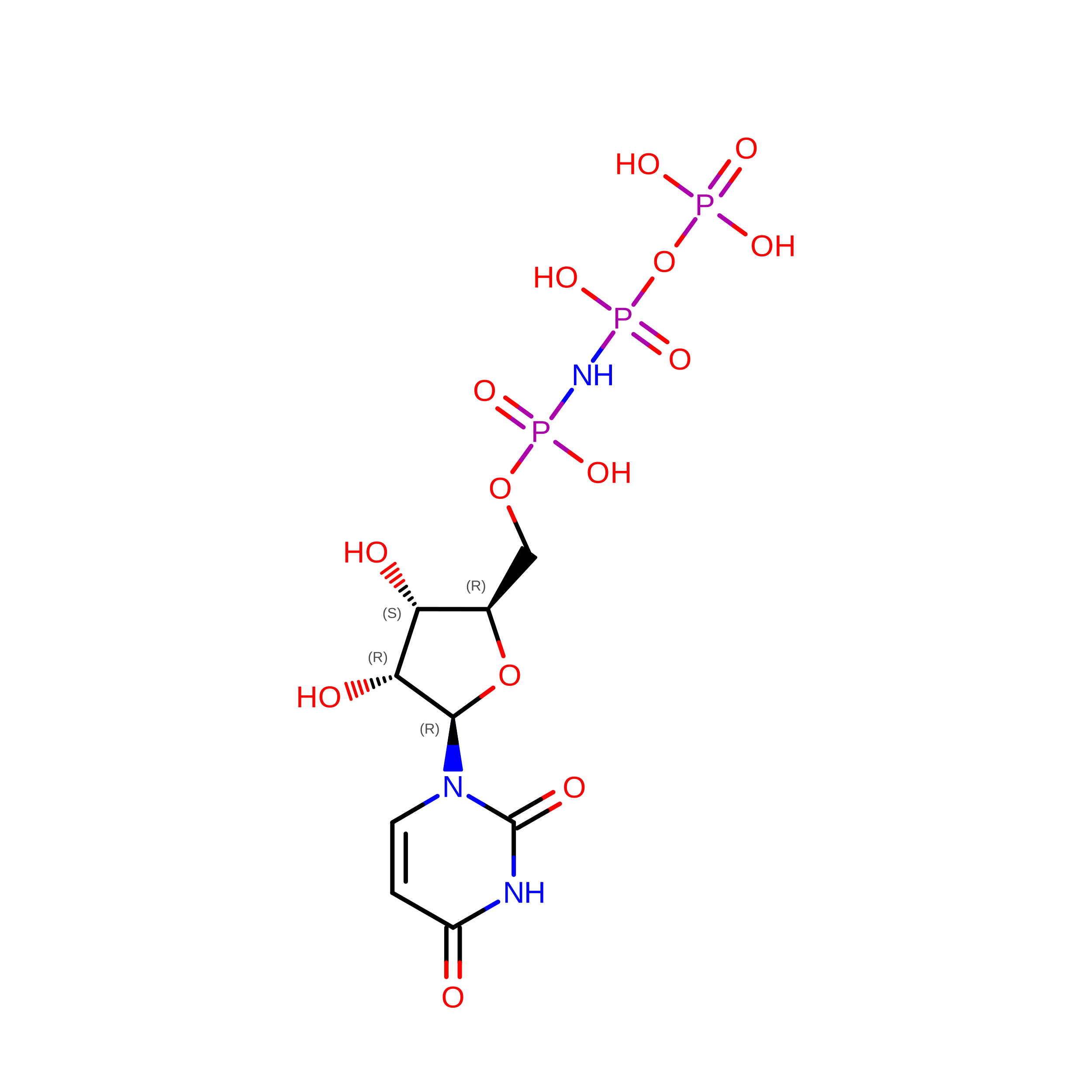
F [auth L]5'-O-[(S)-hydroxy{[(S)-hydroxy(phosphonooxy)phosphoryl]amino}phosphoryl]uridine
C9 H16 N3 O14 P3
OZIBFYOFLVBDIY-XVFCMESISA-N
ZN

Query on ZN


Download:Ideal Coordinates CCD File
L
ZINC ION
Zn
PTFCDOFLOPIGGS-UHFFFAOYSA-N
MN

Query on MN


Download:Ideal Coordinates CCD File
G [auth L],
H [auth L],
I [auth L],
J [auth L],
K [auth L]
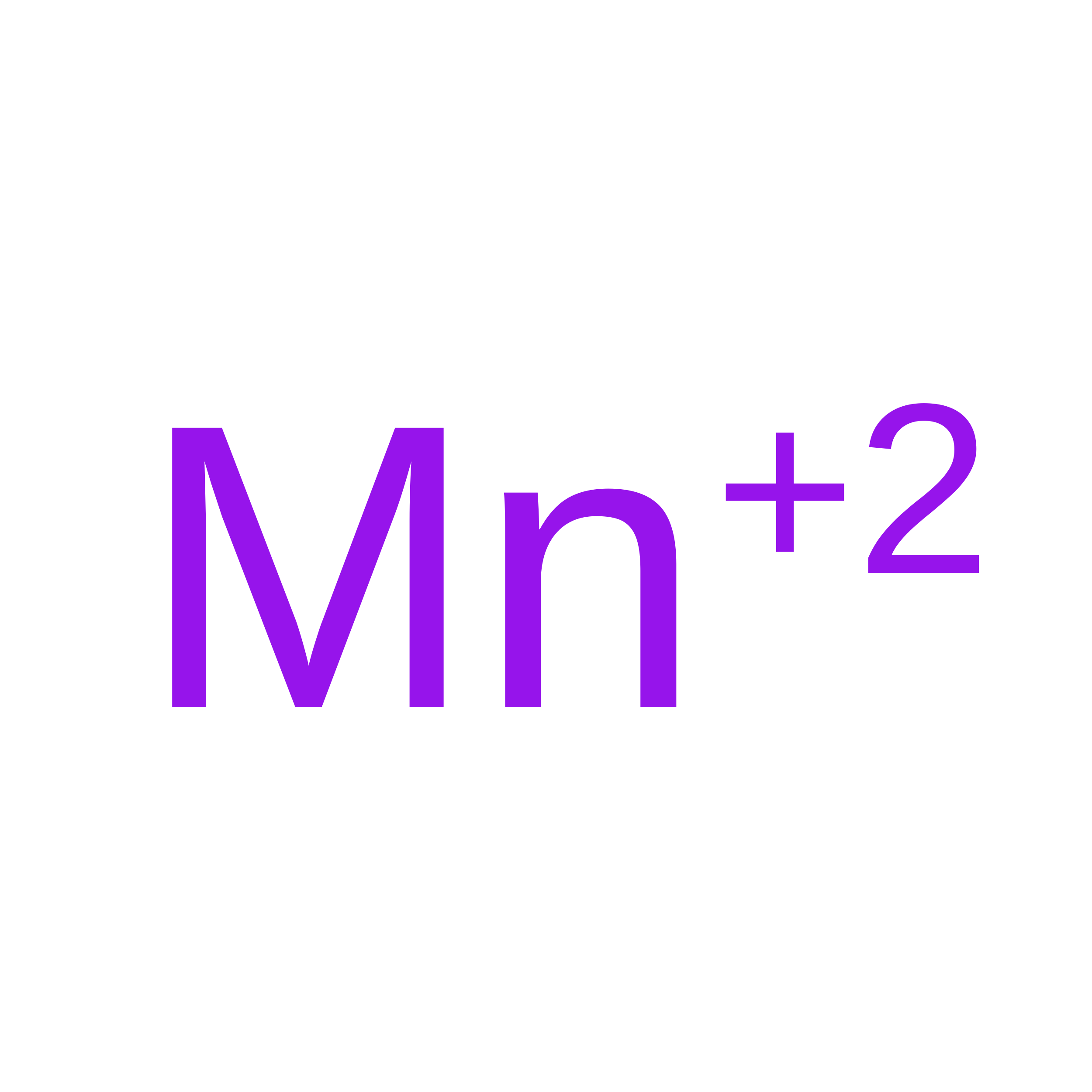
MANGANESE (II) ION
Mn
WAEMQWOKJMHJLA-UHFFFAOYSA-N

Experimental Data & Validation

Experimental Data

  • Method: ELECTRON MICROSCOPY
  • Resolution: 2.92 Å
  • Aggregation State: PARTICLE 
  • Reconstruction Method: SINGLE PARTICLE 
EM Software:
TaskSoftware PackageVersion
MODEL REFINEMENTPHENIX

Structure Validation

View Full Validation Report


Entry History 

& Funding Information

Deposition Data


Funding OrganizationLocationGrant Number
Leibniz AssociationGermanyK27/2017
German Research Foundation (DFG)GermanyINST 152/777-1 FUGG

Revision History  (Full details and data files)

  • Version 1.0: 2021-12-01
    Type: Initial release
  • Version 1.1: 2021-12-22
    Changes: Database references
  • Version 1.2: 2024-05-01
    Changes: Data collection, Structure summary
  • Version 1.3: 2025-07-02
    Changes: Data collection, Structure summary