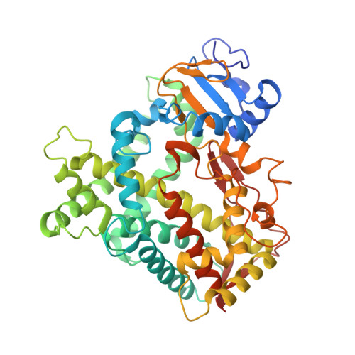
7RL2

Crystal Structure of the Human Cytochrome P450 2C9*8 (CYP2C9*8) Genetic Variant in Complex with the Drug Losartan


Experimental Data Snapshot

  • Method: X-RAY DIFFRACTION
  • Resolution: 2.23 Å
  • R-Value Free: 0.224 
  • R-Value Work: 0.178 
  • R-Value Observed: 0.180 

Starting Model: experimental
View more details

wwPDB Validation   3D Report Full Report


Ligand Structure Quality Assessment 


This is version 1.2 of the entry. See complete history


Literature

Insights into the Genetic Variations of Human Cytochrome P450 2C9: Structural Analysis, Characterization and Comparison.

Parikh, S.J.Kamat, S.Phillips, M.Boyson, S.P.Yarbrough, T.Davie, D.Zhang, Q.Glass, K.C.Shah, M.B.

(2021) Int J Mol Sci 22

  • DOI: https://doi.org/10.3390/ijms221910206
  • Primary Citation of Related Structures:  
    7RL2

  • PubMed Abstract: 

    Cytochromes P450 (CYP) are one of the major xenobiotic metabolizing enzymes with increasing importance in pharmacogenetics. The CYP2C9 enzyme is responsible for the metabolism of a wide range of clinical drugs. More than sixty genetic variations have been identified in CYP2C9 with many demonstrating reduced activity compared to the wild-type (WT) enzyme. The CYP2C9*8 allele is predominantly found in persons of African ancestry and results in altered clearance of several drug substrates of CYP2C9. The X-ray crystal structure of CYP2C9*8, which represents an amino acid variation from arginine to histidine at position 150 (R150H), was solved in complex with losartan. The overall conformation of the CYP2C9*8-losartan complex was similar to the previously solved complex with wild type (WT) protein, but it differs in the occupancy of losartan. One molecule of losartan was bound in the active site and another on the surface in an identical orientation to that observed in the WT complex. However, unlike the WT structure, the losartan in the access channel was not observed in the *8 complex. Furthermore, isothermal titration calorimetry studies illustrated weaker binding of losartan to *8 compared to WT. Interestingly, the CYP2C9*8 interaction with losartan was not as weak as the CYP2C9*3 variant, which showed up to three-fold weaker average dissociation constant compared to the WT. Taken together, the structural and solution characterization yields insights into the similarities and differences of losartan binding to CYP2C9 variants and provides a useful framework for probing the role of amino acid substitution and substrate dependent activity.


  • Organizational Affiliation

    Department of Pharmaceutical Sciences, Albany College of Pharmacy and Health Sciences, 106 New Scotland Avenue, Albany, NY 12208, USA.


Macromolecules
Find similar proteins by:  (by identity cutoff)  |  3D Structure
Entity ID: 1
MoleculeChains Sequence LengthOrganismDetailsImage
Cytochrome P450 2C9476Homo sapiensMutation(s): 2 
Gene Names: CYP2C9CYP2C10
EC: 1.14.14.1 (PDB Primary Data), 1.14.14.53 (PDB Primary Data), 1.14.14.51 (PDB Primary Data), 1.14.14.52 (PDB Primary Data)
Membrane Entity: Yes 
UniProt & NIH Common Fund Data Resources
Find proteins for P11712 (Homo sapiens)
Explore P11712 
Go to UniProtKB:  P11712
PHAROS:  P11712
GTEx:  ENSG00000138109 
Entity Groups  
Sequence Clusters30% Identity50% Identity70% Identity90% Identity95% Identity100% Identity
UniProt GroupP11712
Sequence Annotations
Expand
  • Reference Sequence
Small Molecules
Ligands 4 Unique
IDChains Name / Formula / InChI Key2D Diagram3D Interactions
HEM
Query on HEM

Download Ideal Coordinates CCD File 
B [auth A]PROTOPORPHYRIN IX CONTAINING FE
C34 H32 Fe N4 O4
KABFMIBPWCXCRK-RGGAHWMASA-L
LSN (Subject of Investigation/LOI)
Query on LSN

Download Ideal Coordinates CCD File 
C [auth A],
D [auth A]
[2-butyl-5-chloranyl-3-[[4-[2-(2H-1,2,3,4-tetrazol-5-yl)phenyl]phenyl]methyl]imidazol-4-yl]methanol
C22 H23 Cl N6 O
PSIFNNKUMBGKDQ-UHFFFAOYSA-N
PO4
Query on PO4

Download Ideal Coordinates CCD File 
F [auth A]PHOSPHATE ION
O4 P
NBIIXXVUZAFLBC-UHFFFAOYSA-K
K
Query on K

Download Ideal Coordinates CCD File 
E [auth A]POTASSIUM ION
K
NPYPAHLBTDXSSS-UHFFFAOYSA-N
Experimental Data & Validation

Experimental Data

  • Method: X-RAY DIFFRACTION
  • Resolution: 2.23 Å
  • R-Value Free: 0.224 
  • R-Value Work: 0.178 
  • R-Value Observed: 0.180 
  • Space Group: I 2 2 2
Unit Cell:
Length ( Å )Angle ( ˚ )
a = 75α = 90
b = 143.211β = 90
c = 163.821γ = 90
Software Package:
Software NamePurpose
REFMACrefinement
SCALAdata scaling
PDB_EXTRACTdata extraction
iMOSFLMdata reduction
BALBESphasing

Structure Validation

View Full Validation Report


Ligand Structure Quality Assessment 


Entry History & Funding Information

Deposition Data

  • Released Date: 2021-10-06 
  • Deposition Author(s): Shah, M.B.

Funding OrganizationLocationGrant Number
Not funded--

Revision History  (Full details and data files)

  • Version 1.0: 2021-10-06
    Type: Initial release
  • Version 1.1: 2021-10-27
    Changes: Database references
  • Version 1.2: 2023-10-18
    Changes: Data collection, Refinement description