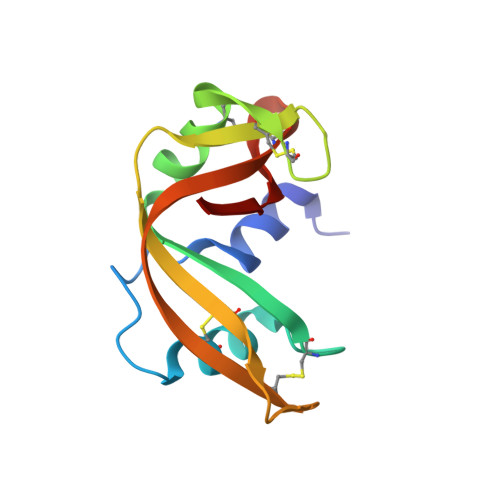
Structure of phosphate-free ribonuclease A refined at 1.26 A.
Wlodawer, A., Svensson, L.A., Sjolin, L., Gilliland, G.L.(1988) Biochemistry 27: 2705-2717
- PubMed: 3401445
- DOI: https://doi.org/10.1021/bi00408a010
- Primary Citation of Related Structures:
7RSA - PubMed Abstract:
The structure of phosphate-free bovine ribonuclease A has been refined at 1.26-A resolution by a restrained least-squares procedure to a final R factor of 0.15. X-ray diffraction data were collected with an electronic position-sensitive detector. The final model consists of all atoms in the polypeptide chain including hydrogens, 188 water sites with full or partial occupancy, and a single molecule of 2-methyl-2-propanol. Thirteen side chains were modeled with two alternate conformations. Major changes to the active site include the addition of two waters in the phosphate-binding pocket, disordering of Gln-11, and tilting of the imidazole ring of His-119. The structure of the protein and of the associated solvent was extensively compared with three other high-resolution, refined structures of this enzyme.
- Center for Chemical Physics, National Bureau of Standards, Gaithersburg, Maryland 20899.
Organizational Affiliation:


