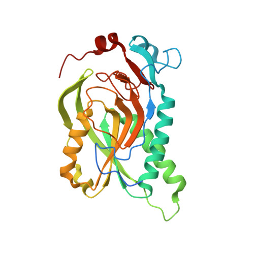
7TUX

Crystal Structure of Plasmodium falciparum Hypoxanthine-Guanine-Xanthine Phosphoribosyltransferase in complex with [(3S)-4-Hydroxy-3-[({2-amino-4-hydroxy-5H-pyrrolo[3,2-d]pyrimidin-7-yl}methyl)amino]butyl]phosphonic acid


Experimental Data Snapshot

  • Method: X-RAY DIFFRACTION
  • Resolution: 1.62 Å
  • R-Value Free: 0.195 
  • R-Value Work: 0.170 
  • R-Value Observed: 0.171 

Starting Model: experimental
View more details

wwPDB Validation   3D Report Full Report


Ligand Structure Quality Assessment 


This is version 1.2 of the entry. See complete history


Literature

Inhibition and Mechanism of Plasmodium falciparum Hypoxanthine-Guanine-Xanthine Phosphoribosyltransferase.

V T Minnow, Y.Suthagar, K.Clinch, K.Ducati, R.G.Ghosh, A.Buckler, J.N.Harijan, R.K.Cahill, S.M.Tyler, P.C.Schramm, V.L.

(2022) ACS Chem Biol 17: 3407-3419

  • DOI: https://doi.org/10.1021/acschembio.2c00546
  • Primary Citation of Related Structures:  
    7TUX

  • PubMed Abstract: 

    Plasmodium falciparum hypoxanthine-guanine-xanthine phosphoribosyltransferase ( Pf HGXPRT) is essential for purine salvage of hypoxanthine into parasite purine nucleotides. Transition state analogue inhibitors of Pf HGXPRT are characterized by kinetic analysis, thermodynamic parameters, and X-ray crystal structures. Compound 1 , 9-deazaguanine linked to an acyclic ribocation phosphonate mimic, shows a kinetic K i of 0.5 nM. Isothermal titration calorimetry (ITC) experiments of 1 binding to Pf HGXPRT reveal enthalpically driven binding with negative cooperativity for the binding of two inhibitor molecules in the tetrameric enzyme. Crystal structures of 1 bound to Pf HGXPRT define the hydrogen bond and ionic contacts to complement binding thermodynamics. Dynamics of ribosyl transfer from 5-phospho-α-d-ribosyl 1-pyrophosphate (PRPP) to hypoxanthine were examined by 18 O isotope exchange at the bridging phosphoryl oxygen of PRPP pyrophosphate. Rotational constraints or short transition state lifetimes prevent torsional rotation and positional isotope exchange of bridging to nonbridging oxygen in the α-pyrophosphoryl group. Thermodynamic analysis of the transition state analogue and magnesium pyrophosphate binding reveal random and cooperative binding to Pf HGXPRT, unlike the obligatory ordered reaction kinetics reported earlier for substrate kinetics.


  • Organizational Affiliation

    Department of Biochemistry, Albert Einstein College of Medicine, Bronx, New York 10461, United States.


Macromolecules
Find similar proteins by:  (by identity cutoff)  |  3D Structure
Entity ID: 1
MoleculeChains Sequence LengthOrganismDetailsImage
Hypoxanthine-guanine-xanthine phosphoribosyltransferase
A, B, C, D
250Plasmodium falciparumMutation(s): 0 
Gene Names: LACZ
EC: 2.4.2.22 (PDB Primary Data), 2.4.2.8 (PDB Primary Data)
UniProt
Find proteins for P20035 (Plasmodium falciparum (isolate FCR-3 / Gambia))
Explore P20035 
Go to UniProtKB:  P20035
Entity Groups  
Sequence Clusters30% Identity50% Identity70% Identity90% Identity95% Identity100% Identity
UniProt GroupP20035
Sequence Annotations
Expand
  • Reference Sequence
Small Molecules
Ligands 5 Unique
IDChains Name / Formula / InChI Key2D Diagram3D Interactions
YBM (Subject of Investigation/LOI)
Query on YBM

Download Ideal Coordinates CCD File 
E [auth A],
HA [auth C],
R [auth B],
WA [auth D]
[(3S)-3-{[(2-amino-4-hydroxy-5H-pyrrolo[3,2-d]pyrimidin-7-yl)methyl]amino}-4-hydroxybutyl]phosphonic acid
C11 H18 N5 O5 P
WDBHHLGLZFXXDD-ZETCQYMHSA-N
POP
Query on POP

Download Ideal Coordinates CCD File 
F [auth A],
IA [auth C],
S [auth B],
XA [auth D]
PYROPHOSPHATE 2-
H2 O7 P2
XPPKVPWEQAFLFU-UHFFFAOYSA-L
EDO
Query on EDO

Download Ideal Coordinates CCD File 
AA [auth B]
AB [auth D]
BA [auth B]
BB [auth D]
CA [auth B]
AA [auth B],
AB [auth D],
BA [auth B],
BB [auth D],
CA [auth B],
CB [auth D],
DA [auth B],
DB [auth D],
EA [auth B],
FA [auth B],
H [auth A],
I [auth A],
J [auth A],
K [auth A],
KA [auth C],
L [auth A],
LA [auth C],
M [auth A],
MA [auth C],
N [auth A],
NA [auth C],
O [auth A],
OA [auth C],
P [auth A],
PA [auth C],
QA [auth C],
RA [auth C],
SA [auth C],
TA [auth C],
U [auth B],
V [auth B],
W [auth B],
X [auth B],
Y [auth B],
Z [auth B],
ZA [auth D]
1,2-ETHANEDIOL
C2 H6 O2
LYCAIKOWRPUZTN-UHFFFAOYSA-N
ACY
Query on ACY

Download Ideal Coordinates CCD File 
EB [auth D]
FB [auth D]
GA [auth B]
Q [auth A]
UA [auth C]
EB [auth D],
FB [auth D],
GA [auth B],
Q [auth A],
UA [auth C],
VA [auth C]
ACETIC ACID
C2 H4 O2
QTBSBXVTEAMEQO-UHFFFAOYSA-N
MG
Query on MG

Download Ideal Coordinates CCD File 
G [auth A],
JA [auth C],
T [auth B],
YA [auth D]
MAGNESIUM ION
Mg
JLVVSXFLKOJNIY-UHFFFAOYSA-N
Experimental Data & Validation

Experimental Data

  • Method: X-RAY DIFFRACTION
  • Resolution: 1.62 Å
  • R-Value Free: 0.195 
  • R-Value Work: 0.170 
  • R-Value Observed: 0.171 
  • Space Group: C 2 2 21
Unit Cell:
Length ( Å )Angle ( ˚ )
a = 105.437α = 90
b = 111.199β = 90
c = 173.652γ = 90
Software Package:
Software NamePurpose
REFMACrefinement
PDB_EXTRACTdata extraction
MOSFLMdata reduction
Aimlessdata scaling
PHASERphasing

Structure Validation

View Full Validation Report


Ligand Structure Quality Assessment 


Entry History & Funding Information

Deposition Data


Funding OrganizationLocationGrant Number
National Institutes of Health/National Institute Of Allergy and Infectious Diseases (NIH/NIAID)United StatesR01-AI127807

Revision History  (Full details and data files)

  • Version 1.0: 2022-12-07
    Type: Initial release
  • Version 1.1: 2022-12-28
    Changes: Database references
  • Version 1.2: 2023-10-25
    Changes: Data collection, Refinement description