


Structural basis of CBP and EP300 interaction with kinase inhibitors
Schonbrunn, E., Bikowitz, M.To be published.
Experimental Data Snapshot
Starting Model: experimental
View more details
Entity ID: 1 | |||||
|---|---|---|---|---|---|
| Molecule | Chains | Sequence Length | Organism | Details | Image |
| Histone acetyltransferase | 116 | Homo sapiens | Mutation(s): 0 Gene Names: CREBBP EC: 2.3.1.48 (PDB Primary Data), 2.3.1 (UniProt) | 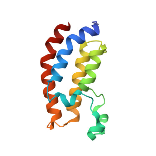 | |
UniProt & NIH Common Fund Data Resources | |||||
PHAROS: Q92793 GTEx: ENSG00000005339 | |||||
Entity Groups | |||||
| Sequence Clusters | 30% Identity50% Identity70% Identity90% Identity95% Identity100% Identity | ||||
| UniProt Group | Q92793 | ||||
Sequence AnnotationsExpand | |||||
Reference Sequence | |||||
| Ligands 2 Unique | |||||
|---|---|---|---|---|---|
| ID | Chains | Name / Formula / InChI Key | 2D Diagram | 3D Interactions | |
| 2LO (Subject of Investigation/LOI) Download:Ideal Coordinates CCD File | D [auth A], F [auth B] | 2-[2-(3-chloro-4-methoxyphenyl)ethyl]-5-(3,5-dimethyl-1,2-oxazol-4-yl)-1-[(2S)-2-(morpholin-4-yl)propyl]-1H-benzimidazole C28 H33 Cl N4 O3 GEPYBHCJBORHCE-SFHVURJKSA-N |  | ||
| N6I (Subject of Investigation/LOI) Download:Ideal Coordinates CCD File | C [auth A], E [auth B] | (3M)-4-{[(2S)-2-(3-chlorophenyl)-2-hydroxyethyl]amino}-3-[4-methyl-6-(morpholin-4-yl)-1H-benzimidazol-2-yl]pyridin-2(1H)-one C25 H26 Cl N5 O3 ZWVZORIKUNOTCS-OAQYLSRUSA-N |  | ||
| Length ( Å ) | Angle ( ˚ ) |
|---|---|
| a = 43.08 | α = 90 |
| b = 59.49 | β = 109.76 |
| c = 57.47 | γ = 90 |
| Software Name | Purpose |
|---|---|
| PHENIX | refinement |
| PDB_EXTRACT | data extraction |
| XDS | data reduction |
| XSCALE | data scaling |
| PHENIX | phasing |
| Funding Organization | Location | Grant Number |
|---|---|---|
| Not funded | -- |


