RCSB PDB - 7UQ2: Vs.4 from T4 phage in complex with cGAMP

 7UQ2

Vs.4 from T4 phage in complex with cGAMP


Experimental Data Snapshot

  • Method: X-RAY DIFFRACTION
  • Resolution: 2.00 Å
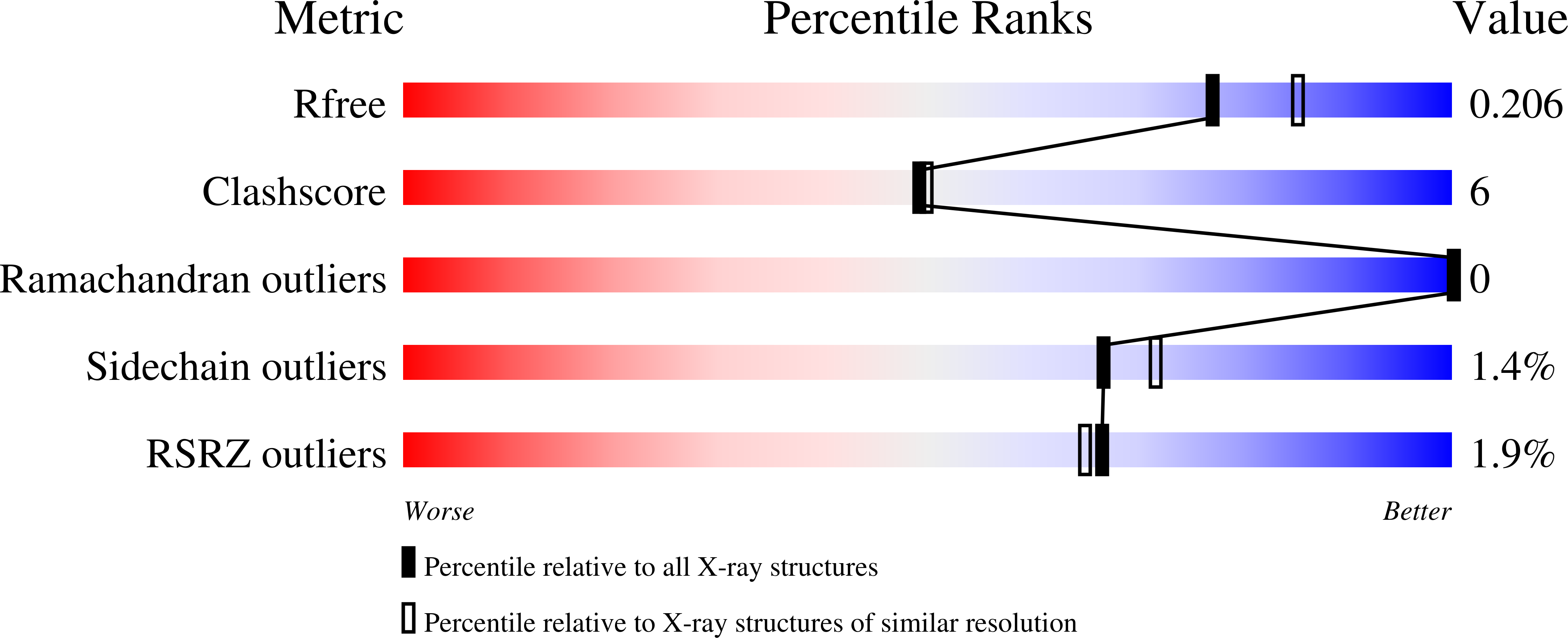
  • R-Value Free: 
    0.206 (Depositor), 0.210 (DCC) 
  • R-Value Work: 
    0.183 (Depositor), 0.180 (DCC) 
  • R-Value Observed: 
    0.184 (Depositor) 

Starting Model: in silico
View more details

wwPDB Validation   3D Report Full Report


Ligand Structure Quality Assessment 

Created with Raphaël 2.3.0Worse 01 BetterLigand structure goodness of fit to experimental dataBest fitted 4BWClick on this verticalbar to view details

This is version 1.4 of the entry. See complete history


Literature

Ubiquitin-like conjugation by bacterial cGAS enhances anti-phage defence.

Jenson, J.M.Li, T.Du, F.Ea, C.K.Chen, Z.J.

(2023) Nature 616: 326-331

  • DOI: https://doi.org/10.1038/s41586-023-05862-7
  • Primary Citation of Related Structures:  
    7UQ2

  • PubMed Abstract: 

    cGAS is an evolutionarily conserved enzyme that has a pivotal role in immune defence against infection 1-3 . In vertebrate animals, cGAS is activated by DNA to produce cyclic GMP-AMP (cGAMP) 4,5 , which leads to the expression of antimicrobial genes 6,7 . In bacteria, cyclic dinucleotide (CDN)-based anti-phage signalling systems (CBASS) have been discovered 8-11 . These systems are composed of cGAS-like enzymes and various effector proteins that kill bacteria on phage infection, thereby stopping phage spread. Of the CBASS systems reported, approximately 39% contain Cap2 and Cap3, which encode proteins with homology to ubiquitin conjugating (E1/E2) and deconjugating enzymes, respectively 8,12 . Although these proteins are required to prevent infection of some bacteriophages 8 , the mechanism by which the enzymatic activities exert an anti-phage effect is unknown. Here we show that Cap2 forms a thioester bond with the C-terminal glycine of cGAS and promotes conjugation of cGAS to target proteins in a process that resembles ubiquitin conjugation. The covalent conjugation of cGAS increases the production of cGAMP. Using a genetic screen, we found that the phage protein Vs.4 antagonized cGAS signalling by binding tightly to cGAMP (dissociation constant of approximately 30 nM) and sequestering it. A crystal structure of Vs.4 bound to cGAMP showed that Vs.4 formed a hexamer that was bound to three molecules of cGAMP. These results reveal a ubiquitin-like conjugation mechanism that regulates cGAS activity in bacteria and illustrates an arms race between bacteria and viruses through controlling CDN levels.


  • Organizational Affiliation

    Department of Molecular Biology, University of Texas Southwestern Medical Center, Dallas, TX, USA.


Macromolecules
Find similar proteins by:  (by identity cutoff)  |  3D Structure
Entity ID: 1
MoleculeChains Sequence LengthOrganismDetailsImage
Vs.4
A, B, C, D, E
89TequatrovirusMutation(s): 0 
Gene Names: y06G62.5vs.4
UniProt
Find proteins for P13314 (Enterobacteria phage T4)
Explore P13314 
Go to UniProtKB:  P13314
Entity Groups  
Sequence Clusters30% Identity50% Identity70% Identity90% Identity95% Identity100% Identity
UniProt GroupP13314
Sequence Annotations
Expand
  • Reference Sequence
Small Molecules
Ligands 2 Unique
IDChains Name / Formula / InChI Key2D Diagram3D Interactions
4BW (Subject of Investigation/LOI)
Query on 4BW

Download Ideal Coordinates CCD File 
G [auth A],
K [auth B],
L [auth C]
2-amino-9-[(2R,3R,3aS,5R,7aR,9R,10R,10aS,12R,14aR)-9-(6-amino-9H-purin-9-yl)-3,5,10,12-tetrahydroxy-5,12-dioxidooctahydro-2H,7H-difuro[3,2-d:3',2'-j][1,3,7,9,2,8]tetraoxadiphosphacyclododecin-2-yl]-1,9-dihydro-6H-purin-6-one
C20 H24 N10 O13 P2
RFCBNSCSPXMEBK-INFSMZHSSA-N
CA
Query on CA

Download Ideal Coordinates CCD File 
H [auth A],
I [auth A],
J [auth A],
M [auth C]
CALCIUM ION
Ca
BHPQYMZQTOCNFJ-UHFFFAOYSA-N
Experimental Data & Validation

Experimental Data

  • Method: X-RAY DIFFRACTION
  • Resolution: 2.00 Å
  • R-Value Free:  0.206 (Depositor), 0.210 (DCC) 
  • R-Value Work:  0.183 (Depositor), 0.180 (DCC) 
  • R-Value Observed: 0.184 (Depositor) 
Space Group: P 41 21 2
Unit Cell:
Length ( Å )Angle ( ˚ )
a = 81.694α = 90
b = 81.694β = 90
c = 217.616γ = 90
Software Package:
Software NamePurpose
PHENIXrefinement
HKL-3000data reduction
HKL-3000data scaling
PHASERphasing

Structure Validation

View Full Validation Report


Ligand Structure Quality Assessment 

Created with Raphaël 2.3.0Worse 01 BetterLigand structure goodness of fit to experimental dataBest fitted 4BWClick on this verticalbar to view details

Entry History & Funding Information

Deposition Data


Funding OrganizationLocationGrant Number
Howard Hughes Medical Institute (HHMI)United States--

Revision History  (Full details and data files)

  • Version 1.0: 2023-02-08
    Type: Initial release
  • Version 1.1: 2023-03-15
    Changes: Database references
  • Version 1.2: 2023-04-26
    Changes: Database references
  • Version 1.3: 2024-04-03
    Changes: Data collection, Refinement description
  • Version 1.4: 2024-10-23
    Changes: Structure summary