7VHC | pdb_00007vhc

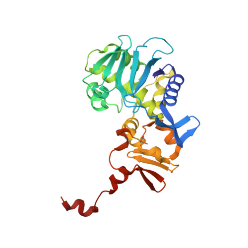
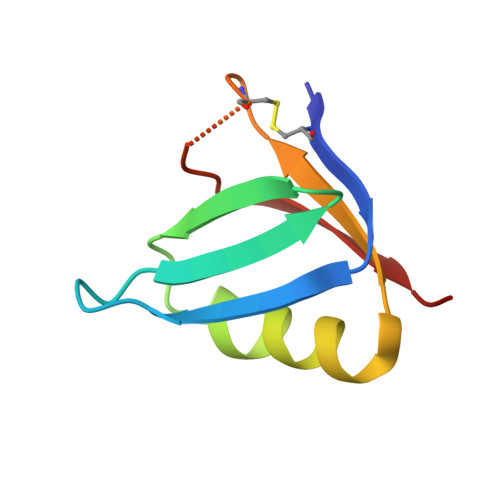
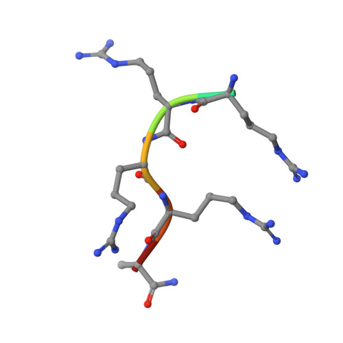
Crystal structure of the STX2a complexed with AR4A peptide


Experimental Data Snapshot

  • Method: X-RAY DIFFRACTION
  • Resolution: 1.80 Å
  • R-Value Free: 
    0.195 (Depositor), 0.195 (DCC) 
  • R-Value Work: 
    0.166 (Depositor), 0.166 (DCC) 
  • R-Value Observed: 
    0.168 (Depositor) 

Starting Model: experimental
View more details

wwPDB Validation 3D Report Full Report

Validation slider image for 7VHC

Ligand Structure Quality Assessment 


This is version 1.2 of the entry. See complete history. 

Literature

A unique peptide-based pharmacophore identifies an inhibitory compound against the A-subunit of Shiga toxin.

Watanabe-Takahashi, M., Senda, M., Yoshino, R., Hibino, M., Hama, S., Terada, T., Shimizu, K., Senda, T., Nishikawa, K.

(2022) Sci Rep 12: 11443-11443

  • DOI: https://doi.org/10.1038/s41598-022-15316-1
  • Primary Citation Related Structures: 
    7VHC, 7VHD, 7VHE, 7VHF

  • PubMed Abstract: 

    Shiga toxin (Stx), a major virulence factor of enterohemorrhagic Escherichia coli (EHEC), can cause fatal systemic complications. Recently, we identified a potent inhibitory peptide that binds to the catalytic A-subunit of Stx. Here, using biochemical structural analysis and X-ray crystallography, we determined a minimal essential peptide motif that occupies the catalytic cavity and is required for binding to the A-subunit of Stx2a, a highly virulent Stx subtype. Molecular dynamics simulations also identified the same motif and allowed determination of a unique pharmacophore for A-subunit binding. Notably, a series of synthetic peptides containing the motif efficiently inhibit Stx2a. In addition, pharmacophore screening and subsequent docking simulations ultimately identified nine Stx2a-interacting molecules out of a chemical compound database consisting of over 7,400,000 molecules. Critically, one of these molecules markedly inhibits Stx2a both in vitro and in vivo, clearly demonstrating the significance of the pharmacophore for identifying therapeutic agents against EHEC infection.


  • Organizational Affiliation: 
    • Department of Molecular Life Sciences, Graduate School of Life and Medical Sciences, Doshisha University, Kyoto, Japan.

Macromolecule Content 

  • Total Structure Weight: 73.94 kDa 
  • Atom Count: 5,530 
  • Modeled Residue Count: 637 
  • Deposited Residue Count: 654 
  • Unique protein chains: 3

Macromolecules

Find similar proteins by:|  3D Structure
Entity ID: 1
MoleculeChains  Sequence LengthOrganismDetailsImage
rRNA N-glycosylase297Escherichia coliMutation(s): 0 
Gene Names: stx2a
EC: 3.2.2.22
UniProt
Find proteins for Q8XBV2 (Escherichia coli)
Explore Q8XBV2 
Go to UniProtKB:  Q8XBV2
Entity Groups
Sequence Clusters30% Identity50% Identity70% Identity90% Identity95% Identity100% Identity
UniProt GroupQ8XBV2
Sequence Annotations
Expand
Reference Sequence
Find similar proteins by:|  3D Structure
Entity ID: 2
MoleculeChains  Sequence LengthOrganismDetailsImage
Shiga toxin 2 B subunit
B, C, D, E, F
70Escherichia coliMutation(s): 0 
Gene Names: stxII, stx2B, stx2B_2, stx2dB, stx2vB, stxB2, vtx2B
UniProt
Find proteins for Q7DJJ2 (Escherichia coli)
Explore Q7DJJ2 
Go to UniProtKB:  Q7DJJ2
Entity Groups
Sequence Clusters30% Identity50% Identity70% Identity90% Identity95% Identity100% Identity
UniProt GroupQ7DJJ2
Sequence Annotations
Expand
Reference Sequence
Find similar proteins by:  Sequence   |   3D Structure  
Entity ID: 3
MoleculeChains  Sequence LengthOrganismDetailsImage
inhibitor peptide, ALA-ARG-ARG-ARG-ARG-ALA7synthetic constructMutation(s): 0 
Sequence Annotations
Expand
Reference Sequence

Experimental Data & Validation

Experimental Data

  • Method: X-RAY DIFFRACTION
  • Resolution: 1.80 Å
  • R-Value Free:  0.195 (Depositor), 0.195 (DCC) 
  • R-Value Work:  0.166 (Depositor), 0.166 (DCC) 
  • R-Value Observed: 0.168 (Depositor) 
Space Group: P 61
Unit Cell:
Length ( Å )Angle ( ˚ )
a = 146.226α = 90
b = 146.226β = 90
c = 60.205γ = 120
Software Package:
Software NamePurpose
PHENIXrefinement
XDSdata reduction
Aimlessdata scaling
MOLREPphasing
Cootmodel building

Structure Validation

View Full Validation Report


Ligand Structure Quality Assessment 


Entry History 

& Funding Information

Deposition Data


Funding OrganizationLocationGrant Number
Japan Agency for Medical Research and Development (AMED)JapanJP20am0101071

Revision History  (Full details and data files)

  • Version 1.0: 2022-07-20
    Type: Initial release
  • Version 1.1: 2023-11-29
    Changes: Data collection, Refinement description
  • Version 1.2: 2024-11-20
    Changes: Structure summary