7VNL

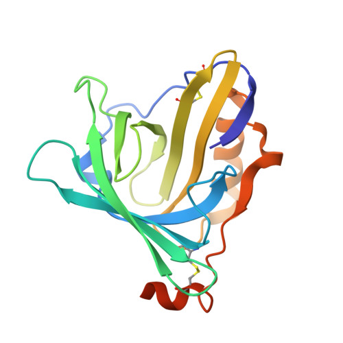
Sandercyanin mutant-F55A-Biliverdin complex


Experimental Data Snapshot

  • Method: X-RAY DIFFRACTION
  • Resolution: 1.93 Å
  • R-Value Free: 0.225 
  • R-Value Work: 0.193 
  • R-Value Observed: 0.195 

Starting Model: experimental
View more details

wwPDB Validation   3D Report Full Report


Ligand Structure Quality Assessment 


This is version 1.2 of the entry. See complete history


Literature

Phenylalanine stacking enhances the red fluorescence of biliverdin IX alpha on UV excitation in sandercyanin fluorescent protein.

Yadav, K.Ghosh, S.Barak, A.Schaefer, W.Subramanian, R.

(2022) FEBS Lett 596: 796-805

  • DOI: https://doi.org/10.1002/1873-3468.14281
  • Primary Citation of Related Structures:  
    7VNL, 7VNS

  • PubMed Abstract: 

    Biliverdin IXα (BV) binds to several prokaryotic and eukaryotic proteins. How nature exploits the versatility of BV's properties is not fully understood. Unlike free BV, the Sandercyanin fluorescent protein bound to BV (SFP-BV) shows enhanced red fluorescence (675 nm) on excitation in the UV region (380 nm). Site-directed mutagenesis showed that the BV complex of two SFP variants, F55A and E79A, resulted in the loss of red fluorescence. Crystal structures of the complexes of these proteins with BV show the absence of stacking interactions of the F55 phenyl ring with BV. BV changes from ZZZssa conformation in the wild-type to ZZZsss conformation in the variants. In the nonfluorescent mutants, the lowest excited state is destabilized, resulting in nonradiative decay.


  • Organizational Affiliation

    Institute for Stem Cell Science and Regenerative Medicine, Bangalore, India.


Macromolecules
Find similar proteins by:  (by identity cutoff)  |  3D Structure
Entity ID: 1
MoleculeChains Sequence LengthOrganismDetailsImage
Sandercyanin Fluorescent Protein
A, B
183Sander vitreusMutation(s): 1 
UniProt
Find proteins for A0A1D5B367 (Sander vitreus)
Explore A0A1D5B367 
Go to UniProtKB:  A0A1D5B367
Entity Groups  
Sequence Clusters30% Identity50% Identity70% Identity90% Identity95% Identity100% Identity
UniProt GroupA0A1D5B367
Sequence Annotations
Expand
  • Reference Sequence
Experimental Data & Validation

Experimental Data

  • Method: X-RAY DIFFRACTION
  • Resolution: 1.93 Å
  • R-Value Free: 0.225 
  • R-Value Work: 0.193 
  • R-Value Observed: 0.195 
  • Space Group: P 63 2 2
Unit Cell:
Length ( Å )Angle ( ˚ )
a = 158.388α = 90
b = 158.388β = 90
c = 81.073γ = 120
Software Package:
Software NamePurpose
PHENIXrefinement
SCALAdata scaling
PHENIXphasing
DIALSdata reduction

Structure Validation

View Full Validation Report


Ligand Structure Quality Assessment 


Entry History & Funding Information

Deposition Data


Funding OrganizationLocationGrant Number
Department of Biotechnology (DBT, India)IndiaBT/PR5801/INF/22/156/2012

Revision History  (Full details and data files)

  • Version 1.0: 2022-05-25
    Type: Initial release
  • Version 1.1: 2023-11-29
    Changes: Data collection, Refinement description
  • Version 1.2: 2024-10-30
    Changes: Structure summary