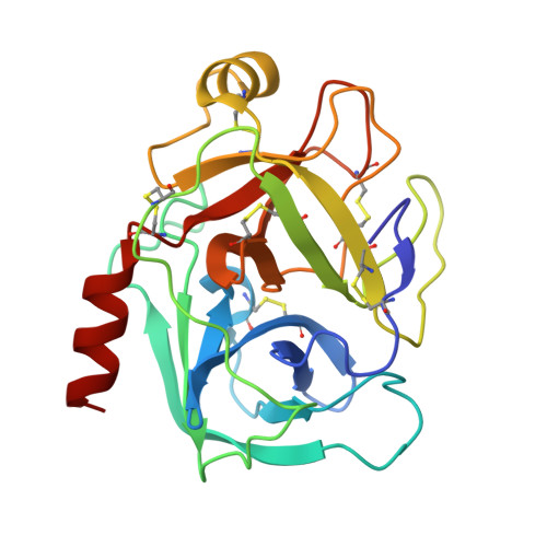
7WBA

Crystal structure of Bovine Pancreatic Trypsin in complex with Tryptamine at Room Temperature


Experimental Data Snapshot

  • Method: X-RAY DIFFRACTION
  • Resolution: 1.45 Å
  • R-Value Free: 0.173 
  • R-Value Work: 0.135 
  • R-Value Observed: 0.137 

Starting Model: experimental
View more details

wwPDB Validation   3D Report Full Report


Ligand Structure Quality Assessment 


This is version 1.2 of the entry. See complete history


Literature

In situ crystal data-collection and ligand-screening system at SPring-8.

Okumura, H.Sakai, N.Murakami, H.Mizuno, N.Nakamura, Y.Ueno, G.Masunaga, T.Kawamura, T.Baba, S.Hasegawa, K.Yamamoto, M.Kumasaka, T.

(2022) Acta Crystallogr F Struct Biol Commun 78: 241-251

  • DOI: https://doi.org/10.1107/S2053230X22005283
  • Primary Citation of Related Structures:  
    7WA0, 7WA2, 7WB6, 7WB7, 7WB8, 7WB9, 7WBA

  • PubMed Abstract: 

    In situ diffraction data collection using crystallization plates has been utilized for macromolecules to evaluate crystal quality without requiring additional sample treatment such as cryocooling. Although it is difficult to collect complete data sets using this technique due to the mechanical limitation of crystal rotation, recent advances in methods for data collection from multiple crystals have overcome this issue. At SPring-8, an in situ diffraction measurement system was constructed consisting of a goniometer for a plate, an articulated robot and plate storage. Using this system, complete data sets were obtained utilizing the small-wedge measurement method. Combining this system with an acoustic liquid handler to prepare protein-ligand complex crystals by applying fragment compounds to trypsin crystals for in situ soaking, binding was confirmed for seven out of eight compounds. These results show that the system functioned properly to collect complete data for structural analysis and to expand the capability for ligand screening in combination with a liquid dispenser.


  • Organizational Affiliation

    Structural Biology Division, Japan Synchrotron Radiation Research Institute, 1-1-1 Kouto, Sayo-cho, Sayo-gun, Hyogo 679-5198, Japan.


Macromolecules
Find similar proteins by:  (by identity cutoff)  |  3D Structure
Entity ID: 1
MoleculeChains Sequence LengthOrganismDetailsImage
Cationic trypsin223Bos taurusMutation(s): 0 
EC: 3.4.21.4
UniProt
Find proteins for P00760 (Bos taurus)
Explore P00760 
Go to UniProtKB:  P00760
Entity Groups  
Sequence Clusters30% Identity50% Identity70% Identity90% Identity95% Identity100% Identity
UniProt GroupP00760
Sequence Annotations
Expand
  • Reference Sequence
Experimental Data & Validation

Experimental Data

  • Method: X-RAY DIFFRACTION
  • Resolution: 1.45 Å
  • R-Value Free: 0.173 
  • R-Value Work: 0.135 
  • R-Value Observed: 0.137 
  • Space Group: P 21 21 21
Unit Cell:
Length ( Å )Angle ( ˚ )
a = 54.61α = 90
b = 58.14β = 90
c = 67.36γ = 90
Software Package:
Software NamePurpose
REFMACrefinement
XDSdata reduction
XSCALEdata scaling
REFMACphasing

Structure Validation

View Full Validation Report


Ligand Structure Quality Assessment 


Entry History & Funding Information

Deposition Data


Funding OrganizationLocationGrant Number
Not funded--

Revision History  (Full details and data files)

  • Version 1.0: 2022-06-15
    Type: Initial release
  • Version 1.1: 2023-11-29
    Changes: Data collection, Refinement description
  • Version 1.2: 2024-10-16
    Changes: Structure summary