8B0B

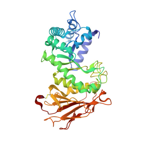
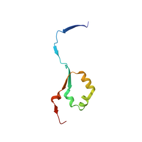
Crystal structure of human heparanase in complex with covalent inhibitor VB151


Experimental Data Snapshot

  • Method: X-RAY DIFFRACTION
  • Resolution: 1.95 Å
  • R-Value Free: 0.291 
  • R-Value Work: 0.232 

Starting Model: experimental
View more details

wwPDB Validation   3D Report Full Report


Ligand Structure Quality Assessment 


This is version 1.4 of the entry. See complete history


Literature

4-O-Substituted Glucuronic Cyclophellitols are Selective Mechanism-Based Heparanase Inhibitors.

Borlandelli, V.Armstrong, Z.Nin-Hill, A.Codee, J.D.C.Raich, L.Artola, M.Rovira, C.Davies, G.J.Overkleeft, H.S.

(2023) ChemMedChem 18: e202200580-e202200580

  • DOI: https://doi.org/10.1002/cmdc.202200580
  • Primary Citation of Related Structures:  
    8B0B, 8B0C, 8B0D, 8B0E

  • PubMed Abstract: 

    Degradation of the extracellular matrix (ECM) supports tissue integrity and homeostasis, but is also a key factor in cancer metastasis. Heparanase (HPSE) is a mammalian ECM-remodeling enzyme with β-D-endo-glucuronidase activity overexpressed in several malignancies, and is thought to facilitate tumor growth and metastasis. By this virtue, HPSE is considered an attractive target for the development of cancer therapies, yet to date no HPSE inhibitors have progressed to the clinic. Here we report on the discovery of glucurono-configured cyclitol derivatives featuring simple substituents at the 4-O-position as irreversible HPSE inhibitors. We show that these compounds, unlike glucurono-cyclophellitol, are selective for HPSE over β-D-exo-glucuronidase (GUSB), also in platelet lysate. The observed selectivity is induced by steric and electrostatic interactions of the substituents at the 4-O-position. Crystallographic analysis supports this rationale for HPSE selectivity, and computer simulations provide insights in the conformational preferences and binding poses of the inhibitors, which we believe are good starting points for the future development of HPSE-targeting antimetastatic cancer drugs.


  • Organizational Affiliation

    Bio-organic Synthesis, Leiden Institute of Chemistry (LIC), Leiden University, Gorlaeus Laboratories, Einsteinweg 55, 2333 CC, Leiden, The Netherlands.


Macromolecules
Find similar proteins by:  (by identity cutoff)  |  3D Structure
Entity ID: 1
MoleculeChains Sequence LengthOrganismDetailsImage
Heparanase 50 kDa subunitA [auth AAA]385Homo sapiensMutation(s): 1 
Gene Names: HPSEHEPHPAHPA1HPR1HPSE1HSE1
EC: 3.2.1.166
UniProt & NIH Common Fund Data Resources
Find proteins for Q9Y251 (Homo sapiens)
Explore Q9Y251 
Go to UniProtKB:  Q9Y251
PHAROS:  Q9Y251
GTEx:  ENSG00000173083 
Entity Groups  
Sequence Clusters30% Identity50% Identity70% Identity90% Identity95% Identity100% Identity
UniProt GroupQ9Y251
Glycosylation
Glycosylation Sites: 3Go to GlyGen: Q9Y251-1
Sequence Annotations
Expand
  • Reference Sequence
Find similar proteins by:  (by identity cutoff)  |  3D Structure
Entity ID: 2
MoleculeChains Sequence LengthOrganismDetailsImage
Heparanase 8 kDa subunitB [auth BBB]74Homo sapiensMutation(s): 0 
Gene Names: HPSEHEPHPAHPA1HPR1HPSE1HSE1
EC: 3.2.1.166
UniProt & NIH Common Fund Data Resources
Find proteins for Q9Y251 (Homo sapiens)
Explore Q9Y251 
Go to UniProtKB:  Q9Y251
PHAROS:  Q9Y251
GTEx:  ENSG00000173083 
Entity Groups  
Sequence Clusters30% Identity50% Identity70% Identity90% Identity95% Identity100% Identity
UniProt GroupQ9Y251
Sequence Annotations
Expand
  • Reference Sequence
Small Molecules
Ligands 3 Unique
IDChains Name / Formula / InChI Key2D Diagram3D Interactions
OUU (Subject of Investigation/LOI)
Query on OUU

Download Ideal Coordinates CCD File 
G [auth AAA](1~{S},2~{R},3~{R},4~{S},6~{S})-2-(2-acetamidoethoxy)-3,4,6-tris(oxidanyl)cyclohexane-1-carboxylic acid
C11 H19 N O7
PCCLVLLYDLIJIM-RXKWGBCNSA-N
NAG
Query on NAG

Download Ideal Coordinates CCD File 
C [auth AAA],
D [auth AAA],
E [auth AAA]
2-acetamido-2-deoxy-beta-D-glucopyranose
C8 H15 N O6
OVRNDRQMDRJTHS-FMDGEEDCSA-N
EDO
Query on EDO

Download Ideal Coordinates CCD File 
F [auth AAA]1,2-ETHANEDIOL
C2 H6 O2
LYCAIKOWRPUZTN-UHFFFAOYSA-N
Experimental Data & Validation

Experimental Data

  • Method: X-RAY DIFFRACTION
  • Resolution: 1.95 Å
  • R-Value Free: 0.291 
  • R-Value Work: 0.232 
  • Space Group: P 1 21 1
Unit Cell:
Length ( Å )Angle ( ˚ )
a = 44.442α = 90
b = 70.846β = 98.702
c = 78.293γ = 90
Software Package:
Software NamePurpose
REFMACrefinement
DIALSdata reduction
DIALSdata scaling
PHASERphasing

Structure Validation

View Full Validation Report


Ligand Structure Quality Assessment 


Entry History & Funding Information

Deposition Data


Funding OrganizationLocationGrant Number
Biotechnology and Biological Sciences Research Council (BBSRC)United Kingdom--

Revision History  (Full details and data files)

  • Version 1.0: 2022-12-28
    Type: Initial release
  • Version 1.1: 2023-02-01
    Changes: Database references
  • Version 1.2: 2023-03-01
    Changes: Database references
  • Version 1.3: 2024-02-07
    Changes: Data collection, Refinement description
  • Version 1.4: 2024-11-13
    Changes: Structure summary