8BRC

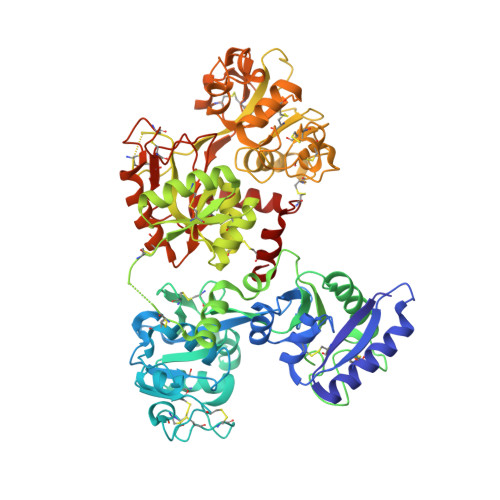
Crystal structure of the adduct between human serum transferrin and cisplatin


Experimental Data Snapshot

  • Method: X-RAY DIFFRACTION
  • Resolution: 3.17 Å
  • R-Value Free: 0.243 
  • R-Value Work: 0.177 
  • R-Value Observed: 0.181 

Starting Model: experimental
View more details

wwPDB Validation   3D Report Full Report


This is version 1.2 of the entry. See complete history


Literature

Cisplatin Binding to Human Serum Transferrin: A Crystallographic Study.

Troisi, R.Galardo, F.Ferraro, G.Sica, F.Merlino, A.

(2023) Inorg Chem 62: 675-678

  • DOI: https://doi.org/10.1021/acs.inorgchem.2c04206
  • Primary Citation of Related Structures:  
    8BRC

  • PubMed Abstract: 

    The molecular mechanism of how human serum transferrin (hTF) recognizes cisplatin at the atomic level is still unclear. Here, we report the molecular structure of the adduct formed upon the reaction of hTF with cisplatin. Pt binds the side chain of Met256 (at the N-lobe), without altering the protein overall conformation.


  • Organizational Affiliation

    Department of Chemical Sciences, University of Naples Federico II, Complesso Universitario di Monte Sant'Angelo, via Cintia, Naples I-80126, Italy.


Macromolecules
Find similar proteins by:  (by identity cutoff)  |  3D Structure
Entity ID: 1
MoleculeChains Sequence LengthOrganismDetailsImage
Serotransferrin679Homo sapiensMutation(s): 0 
UniProt & NIH Common Fund Data Resources
Find proteins for P02787 (Homo sapiens)
Explore P02787 
Go to UniProtKB:  P02787
PHAROS:  P02787
GTEx:  ENSG00000091513 
Entity Groups  
Sequence Clusters30% Identity50% Identity70% Identity90% Identity95% Identity100% Identity
UniProt GroupP02787
Glycosylation
Glycosylation Sites: 1Go to GlyGen: P02787-1
Sequence Annotations
Expand
  • Reference Sequence
Small Molecules
Ligands 5 Unique
IDChains Name / Formula / InChI Key2D Diagram3D Interactions
NAG
Query on NAG

Download Ideal Coordinates CCD File 
D [auth A]2-acetamido-2-deoxy-beta-D-glucopyranose
C8 H15 N O6
OVRNDRQMDRJTHS-FMDGEEDCSA-N
PT (Subject of Investigation/LOI)
Query on PT

Download Ideal Coordinates CCD File 
E [auth A]PLATINUM (II) ION
Pt
HRGDZIGMBDGFTC-UHFFFAOYSA-N
MLI
Query on MLI

Download Ideal Coordinates CCD File 
C [auth A]MALONATE ION
C3 H2 O4
OFOBLEOULBTSOW-UHFFFAOYSA-L
FE
Query on FE

Download Ideal Coordinates CCD File 
B [auth A]FE (III) ION
Fe
VTLYFUHAOXGGBS-UHFFFAOYSA-N
NH3 (Subject of Investigation/LOI)
Query on NH3

Download Ideal Coordinates CCD File 
F [auth A],
G [auth A]
AMMONIA
H3 N
QGZKDVFQNNGYKY-UHFFFAOYSA-N
Experimental Data & Validation

Experimental Data

  • Method: X-RAY DIFFRACTION
  • Resolution: 3.17 Å
  • R-Value Free: 0.243 
  • R-Value Work: 0.177 
  • R-Value Observed: 0.181 
  • Space Group: C 2 2 21
Unit Cell:
Length ( Å )Angle ( ˚ )
a = 136.34α = 90
b = 156.4β = 90
c = 107.44γ = 90
Software Package:
Software NamePurpose
REFMACrefinement
XDSdata reduction
XSCALEdata scaling
PHASERphasing
autoPROCdata processing

Structure Validation

View Full Validation Report


Entry History & Funding Information

Deposition Data


Funding OrganizationLocationGrant Number
Not funded--

Revision History  (Full details and data files)

  • Version 1.0: 2023-01-18
    Type: Initial release
  • Version 1.1: 2023-01-25
    Changes: Database references
  • Version 1.2: 2024-02-07
    Changes: Data collection