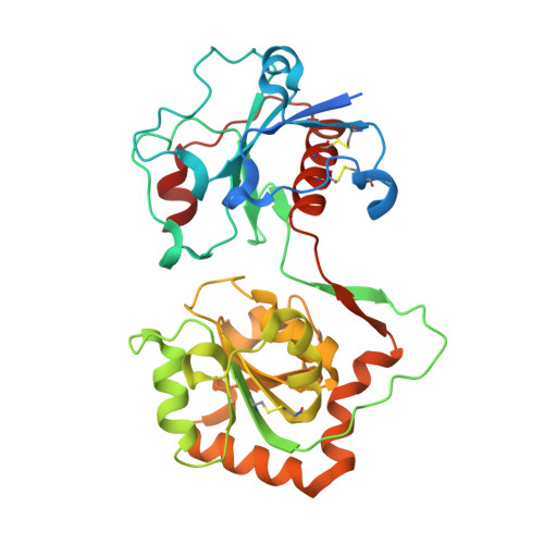
8D0O

Human alpha1,3-fucosyltransferase FUT9, heavy atom derivative

  • Classification: TRANSFERASE
  • Organism(s): Homo sapiens
  • Expression System: Homo sapiens
  • Mutation(s): Yes 

  • Deposited: 2022-05-26 Released: 2023-05-24 
  • Deposition Author(s): Kadirvelraj, R., Wood, Z.A.
  • Funding Organization(s): National Institutes of Health/National Institute on Alcohol Abuse and Alcoholism (NIH/NIAAA)

Experimental Data Snapshot

  • Method: X-RAY DIFFRACTION
  • Resolution: 2.10 Å
  • R-Value Free: 0.209 
  • R-Value Work: 0.164 
  • R-Value Observed: 0.167 

wwPDB Validation   3D Report Full Report


This is version 1.3 of the entry. See complete history


Literature

Structural basis for Lewis antigen synthesis by the alpha 1,3-fucosyltransferase FUT9.

Kadirvelraj, R.Boruah, B.M.Wang, S.Chapla, D.Huang, C.Ramiah, A.Hudson, K.L.Prudden, A.R.Boons, G.J.Withers, S.G.Wood, Z.A.Moremen, K.W.

(2023) Nat Chem Biol 19: 1022-1030

  • DOI: https://doi.org/10.1038/s41589-023-01345-y
  • Primary Citation of Related Structures:  
    8D0O, 8D0P, 8D0Q, 8D0R, 8D0S, 8D0U, 8D0W, 8D0X

  • PubMed Abstract: 

    Mammalian cell surface and secreted glycoproteins exhibit remarkable glycan structural diversity that contributes to numerous physiological and pathogenic interactions. Terminal glycan structures include Lewis antigens synthesized by a collection of α1,3/4-fucosyltransferases (CAZy GT10 family). At present, the only available crystallographic structure of a GT10 member is that of the Helicobacter pylori α1,3-fucosyltransferase, but mammalian GT10 fucosyltransferases are distinct in sequence and substrate specificity compared with the bacterial enzyme. Here, we determined crystal structures of human FUT9, an α1,3-fucosyltransferase that generates Lewis x and Lewis y antigens, in complex with GDP, acceptor glycans, and as a FUT9-donor analog-acceptor Michaelis complex. The structures reveal substrate specificity determinants and allow prediction of a catalytic model supported by kinetic analyses of numerous active site mutants. Comparisons with other GT10 fucosyltransferases and GT-B fold glycosyltransferases provide evidence for modular evolution of donor- and acceptor-binding sites and specificity for Lewis antigen synthesis among mammalian GT10 fucosyltransferases.


  • Organizational Affiliation

    Department of Biochemistry & Molecular Biology, University of Georgia, Athens, GA, USA.


Macromolecules
Find similar proteins by:  (by identity cutoff)  |  3D Structure
Entity ID: 1
MoleculeChains Sequence LengthOrganismDetailsImage
4-galactosyl-N-acetylglucosaminide 3-alpha-L-fucosyltransferase 9322Homo sapiensMutation(s): 1 
Gene Names: FUT9
EC: 2.4.1.152
UniProt & NIH Common Fund Data Resources
Find proteins for Q9Y231 (Homo sapiens)
Explore Q9Y231 
Go to UniProtKB:  Q9Y231
PHAROS:  Q9Y231
GTEx:  ENSG00000172461 
Entity Groups  
Sequence Clusters30% Identity50% Identity70% Identity90% Identity95% Identity100% Identity
UniProt GroupQ9Y231
Glycosylation
Glycosylation Sites: 1Go to GlyGen: Q9Y231-1
Sequence Annotations
Expand
  • Reference Sequence
Oligosaccharides

Help

Entity ID: 2
MoleculeChains Length2D Diagram Glycosylation3D Interactions
2-acetamido-2-deoxy-beta-D-glucopyranose-(1-4)-2-acetamido-2-deoxy-beta-D-glucopyranose
B
2N-Glycosylation
Glycosylation Resources
GlyTouCan:  G42666HT
GlyCosmos:  G42666HT
GlyGen:  G42666HT
Small Molecules
Ligands 2 Unique
IDChains Name / Formula / InChI Key2D Diagram3D Interactions
CS (Subject of Investigation/LOI)
Query on CS

Download Ideal Coordinates CCD File 
Q [auth A]
R [auth A]
S [auth A]
T [auth A]
U [auth A]
Q [auth A],
R [auth A],
S [auth A],
T [auth A],
U [auth A],
V [auth A],
W [auth A],
X [auth A],
Y [auth A]
CESIUM ION
Cs
NCMHKCKGHRPLCM-UHFFFAOYSA-N
IOD (Subject of Investigation/LOI)
Query on IOD

Download Ideal Coordinates CCD File 
C [auth A]
D [auth A]
E [auth A]
F [auth A]
G [auth A]
C [auth A],
D [auth A],
E [auth A],
F [auth A],
G [auth A],
H [auth A],
I [auth A],
J [auth A],
K [auth A],
L [auth A],
M [auth A],
N [auth A],
O [auth A],
P [auth A]
IODIDE ION
I
XMBWDFGMSWQBCA-UHFFFAOYSA-M
Experimental Data & Validation

Experimental Data

  • Method: X-RAY DIFFRACTION
  • Resolution: 2.10 Å
  • R-Value Free: 0.209 
  • R-Value Work: 0.164 
  • R-Value Observed: 0.167 
  • Space Group: I 21 3
Unit Cell:
Length ( Å )Angle ( ˚ )
a = 127.12α = 90
b = 127.12β = 90
c = 127.12γ = 90
Software Package:
Software NamePurpose
PHENIXrefinement
XDSdata reduction
XSCALEdata scaling
PHASERphasing

Structure Validation

View Full Validation Report


Entry History & Funding Information

Deposition Data


Funding OrganizationLocationGrant Number
National Institutes of Health/National Institute on Alcohol Abuse and Alcoholism (NIH/NIAAA)United StatesGM130915, GM103390 and P01GM107012

Revision History  (Full details and data files)

  • Version 1.0: 2023-05-24
    Type: Initial release
  • Version 1.1: 2023-06-07
    Changes: Database references
  • Version 1.2: 2023-08-09
    Changes: Database references
  • Version 1.3: 2024-10-16
    Changes: Data collection, Structure summary