8D5A | pdb_00008d5a

Middle state of SARS-CoV-2 BA.2 variant spike protein


Experimental Data Snapshot

  • Method: ELECTRON MICROSCOPY
  • Resolution: 3.10 Å
  • Aggregation State: PARTICLE 
  • Reconstruction Method: SINGLE PARTICLE 

Starting Model: experimental
View more details

wwPDB Validation 3D Report Full Report

Validation slider image for 8D5A

This is version 1.3 of the entry. See complete history

Literature

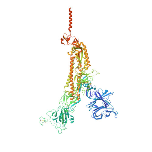
Structural and functional characteristics of the SARS-CoV-2 Omicron subvariant BA.2 spike protein.

Zhang, J.Tang, W.Gao, H.Lavine, C.L.Shi, W.Peng, H.Zhu, H.Anand, K.Kosikova, M.Kwon, H.J.Tong, P.Gautam, A.Rits-Volloch, S.Wang, S.Mayer, M.L.Wesemann, D.R.Seaman, M.S.Lu, J.Xiao, T.Xie, H.Chen, B.

(2023) Nat Struct Mol Biol 30: 980-990

  • DOI: https://doi.org/10.1038/s41594-023-01023-6
  • Primary Citation Related Structures: 
    8D55, 8D56, 8D5A

  • PubMed Abstract: 

    The Omicron subvariant BA.2 has become the dominant circulating strain of severe acute respiratory syndrome coronavirus 2 (SARS-CoV-2) in many countries. Here, we have characterized structural, functional and antigenic properties of the full-length BA.2 spike (S) protein and compared replication of the authentic virus in cell culture and an animal model with previously prevalent variants. BA.2 S can fuse membranes slightly more efficiently than Omicron BA.1, but still less efficiently than other previous variants. Both BA.1 and BA.2 viruses replicated substantially faster in animal lungs than the early G614 (B.1) strain in the absence of pre-existing immunity, possibly explaining the increased transmissibility despite their functionally compromised spikes. As in BA.1, mutations in the BA.2 S remodel its antigenic surfaces, leading to strong resistance to neutralizing antibodies. These results suggest that both immune evasion and replicative advantage may contribute to the heightened transmissibility of the Omicron subvariants.


  • Organizational Affiliation
    • Division of Molecular Medicine, Boston Children's Hospital, Boston, MA, USA.

Macromolecule Content 

  • Total Structure Weight: 454.15 kDa 
  • Atom Count: 27,711 
  • Modeled Residue Count: 3,365 
  • Deposited Residue Count: 3,915 
  • Unique protein chains: 1

Macromolecules

Find similar proteins by:|  3D Structure
Entity ID: 1
MoleculeChains  Sequence LengthOrganismDetailsImage
Spike glycoprotein
A, B, C
1,305Severe acute respiratory syndrome coronavirus 2Mutation(s): 28 
Gene Names: S2
UniProt
Find proteins for P0DTC2 (Severe acute respiratory syndrome coronavirus 2)
Explore P0DTC2 
Go to UniProtKB:  P0DTC2
Entity Groups
Sequence Clusters30% Identity50% Identity70% Identity90% Identity95% Identity100% Identity
UniProt GroupP0DTC2
Glycosylation
Glycosylation Sites: 16Go to GlyGen: P0DTC2-1
Sequence Annotations
Expand
Reference Sequence

Oligosaccharides

Help  
Entity ID: 3
MoleculeChains Length2D Diagram GlycosylationD Interactions
2-acetamido-2-deoxy-beta-D-glucopyranose-(1-4)-2-acetamido-2-deoxy-beta-D-glucopyranose
AA [auth a],
E,
G,
N,
O,
AA [auth a],
E,
G,
N,
O,
Q,
X,
Y
2N-Glycosylation
Glycosylation Resources
GlyTouCan: G42666HT
GlyCosmos: G42666HT
GlyGen: G42666HT
Entity ID: 4
MoleculeChains Length2D Diagram GlycosylationD Interactions
2-acetamido-2-deoxy-beta-D-glucopyranose-(1-4)-[alpha-L-fucopyranose-(1-6)]2-acetamido-2-deoxy-beta-D-glucopyranoseDA [auth d],
J,
T
3N-Glycosylation
Glycosylation Resources
GlyTouCan: G21290RB
GlyCosmos: G21290RB
GlyGen: G21290RB

Small Molecules

Ligands 1 Unique
IDChains Name / Formula / InChI Key2D Diagram3D Interactions
NAG
(Subject of Investigation/LOI)

Query on NAG


Download:Ideal Coordinates CCD File
GA [auth A]
HA [auth A]
IA [auth A]
JA [auth A]
KA [auth A]
GA [auth A],
HA [auth A],
IA [auth A],
JA [auth A],
KA [auth A],
LA [auth A],
MA [auth A],
NA [auth B],
OA [auth B],
PA [auth B],
QA [auth B],
RA [auth B],
SA [auth B],
TA [auth B],
UA [auth C],
VA [auth C],
WA [auth C],
XA [auth C],
YA [auth C],
ZA [auth C]
2-acetamido-2-deoxy-beta-D-glucopyranose
C8 H15 N O6
OVRNDRQMDRJTHS-FMDGEEDCSA-N

Experimental Data & Validation

Experimental Data

  • Method: ELECTRON MICROSCOPY
  • Resolution: 3.10 Å
  • Aggregation State: PARTICLE 
  • Reconstruction Method: SINGLE PARTICLE 
EM Software:
TaskSoftware PackageVersion
MODEL REFINEMENTPHENIX
RECONSTRUCTIONRELION
RECONSTRUCTIONcryoSPARC

Structure Validation

View Full Validation Report


Entry History 

& Funding Information

Deposition Data


Funding OrganizationLocationGrant Number
National Institutes of Health/National Institute Of Allergy and Infectious Diseases (NIH/NIAID)United StatesAI147884
National Institutes of Health/National Institute Of Allergy and Infectious Diseases (NIH/NIAID)United StatesAI141002
National Institutes of Health/National Institute Of Allergy and Infectious Diseases (NIH/NIAID)United StatesAI127193
National Institutes of Health/National Institute Of Allergy and Infectious Diseases (NIH/NIAID)United StatesAI39538
National Institutes of Health/National Institute Of Allergy and Infectious Diseases (NIH/NIAID)United StatesAI165072
National Institutes of Health/National Institute Of Allergy and Infectious Diseases (NIH/NIAID)United StatesAI69619

Revision History  (Full details and data files)

  • Version 1.0: 2023-06-07
    Type: Initial release
  • Version 1.1: 2023-07-12
    Changes: Database references, Other
  • Version 1.2: 2023-07-26
    Changes: Database references
  • Version 1.3: 2024-11-06
    Changes: Data collection, Refinement description, Structure summary