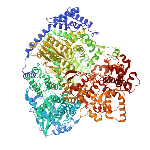
structure of Tomato spotted wilt virus L protein
Cao, L., Wang, X.To be published.
Experimental Data Snapshot
wwPDB Validation 3D Report Full Report
Entity ID: 1 | |||||
|---|---|---|---|---|---|
| Molecule | Chains | Sequence Length | Organism | Details | Image |
| RNA-directed RNA polymerase L | 1,775 | Orthotospovirus tomatomaculae | Mutation(s): 0 EC: 2.7.7.48 |  | |
UniProt | |||||
Find proteins for A0A7G8JUR3 (Tomato spotted wilt virus) Explore A0A7G8JUR3 Go to UniProtKB: A0A7G8JUR3 | |||||
Entity Groups | |||||
| Sequence Clusters | 30% Identity50% Identity70% Identity90% Identity95% Identity100% Identity | ||||
| UniProt Group | A0A7G8JUR3 | ||||
Sequence AnnotationsExpand | |||||
| |||||
Find similar nucleic acids by: Sequence | 3D Structure
Entity ID: 2 | |||||
|---|---|---|---|---|---|
| Molecule | Chains | Length | Organism | Image | |
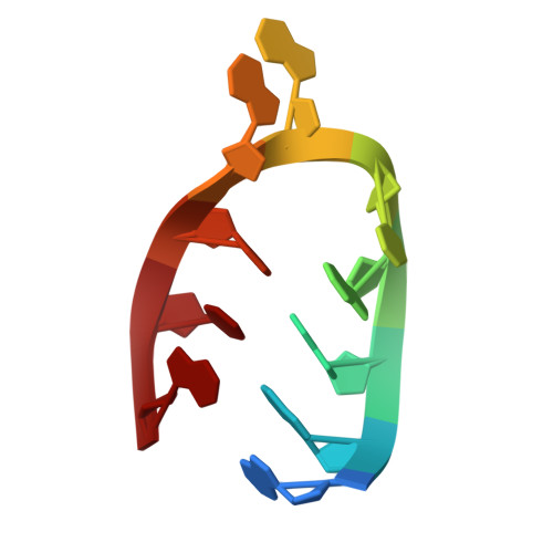
| RNA (5'-R(P*AP*GP*AP*GP*CP*AP*AP*UP*CP*A)-3') | B [auth E] | 10 | synthetic construct |  | |
Sequence AnnotationsExpand | |||||
| |||||
| Funding Organization | Location | Grant Number |
|---|---|---|
| Not funded | -- |