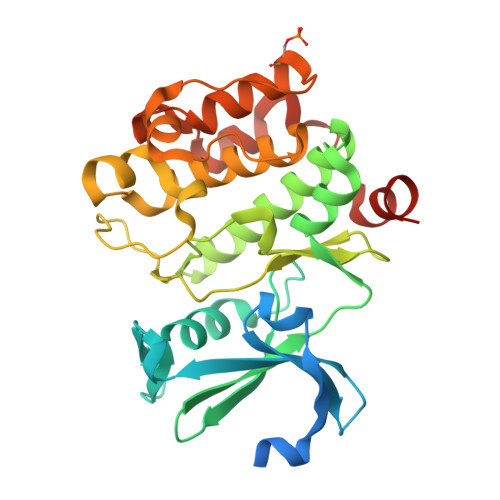
8R18

Pim1 in complex with (E)-4-(4-hydroxystyryl)benzoic acid and Pimtide


Experimental Data Snapshot

  • Method: X-RAY DIFFRACTION
  • Resolution: 1.89 Å
  • R-Value Free: 0.186 
  • R-Value Work: 0.160 
  • R-Value Observed: 0.161 

Starting Model: experimental
View more details

wwPDB Validation   3D Report Full Report


Ligand Structure Quality Assessment 


This is version 1.3 of the entry. See complete history


Literature

What doesn't fit is made to fit: Pim-1 kinase adapts to the configuration of stilbene-based inhibitors.

Hochban, P.M.M.Heyder, L.Heine, A.Diederich, W.E.

(2024) Arch Pharm (Weinheim) 357: e2400094-e2400094

  • DOI: https://doi.org/10.1002/ardp.202400094
  • Primary Citation of Related Structures:  
    8R10, 8R18, 8R1N, 8R1P, 8R1T

  • PubMed Abstract: 

    Recently, we have developed novel Pim-1 kinase inhibitors starting from a dihydrobenzofuran core structure using a computational approach. Here, we report the design and synthesis of stilbene-based Pim-1 kinase inhibitors obtained by formal elimination of the dihydrofuran ring. These inhibitors of the first design cycle, which were obtained as inseparable cis/trans mixtures, showed affinities in the low single-digit micromolar range. To be able to further optimize these compounds in a structure-based fashion, we determined the X-ray structures of the protein-ligand-complexes. Surprisingly, only the cis-isomer binds upon crystallization of the cis/trans-mixture of the ligands with Pim-1 kinase and the substrate PIMTIDE, the binding mode being largely consistent with that predicted by docking. After crystallization of the exclusively trans-configured derivatives, a markedly different binding mode for the inhibitor and a concomitant rearrangement of the glycine-rich loop is observed, resulting in the ligand being deeply buried in the binding pocket.


  • Organizational Affiliation

    Institut für Pharmazeutische Chemie, Zentrum für Tumor und Immunbiologie, Philipps-Universität Marburg, Marburg, Germany.


Macromolecules
Find similar proteins by:  (by identity cutoff)  |  3D Structure
Entity ID: 1
MoleculeChains Sequence LengthOrganismDetailsImage
Serine/threonine-protein kinase pim-1313Homo sapiensMutation(s): 1 
Gene Names: PIM1
EC: 2.7.11.1
UniProt & NIH Common Fund Data Resources
Find proteins for P11309 (Homo sapiens)
Explore P11309 
Go to UniProtKB:  P11309
PHAROS:  P11309
GTEx:  ENSG00000137193 
Entity Groups  
Sequence Clusters30% Identity50% Identity70% Identity90% Identity95% Identity100% Identity
UniProt GroupP11309
Sequence Annotations
Expand
  • Reference Sequence

Find similar proteins by:  Sequence   |   3D Structure  

Entity ID: 2
MoleculeChains Sequence LengthOrganismDetailsImage
PimtideB [auth C]14Homo sapiensMutation(s): 0 
Entity Groups  
Sequence Clusters30% Identity50% Identity70% Identity90% Identity95% Identity100% Identity
Sequence Annotations
Expand
  • Reference Sequence
Experimental Data & Validation

Experimental Data

  • Method: X-RAY DIFFRACTION
  • Resolution: 1.89 Å
  • R-Value Free: 0.186 
  • R-Value Work: 0.160 
  • R-Value Observed: 0.161 
  • Space Group: P 65
Unit Cell:
Length ( Å )Angle ( ˚ )
a = 96.507α = 90
b = 96.507β = 90
c = 80.201γ = 120
Software Package:
Software NamePurpose
PHENIXrefinement
MxCuBEdata collection
XDSdata reduction
XDSdata scaling
PHASERphasing
Cootmodel building

Structure Validation

View Full Validation Report


Ligand Structure Quality Assessment 


Entry History & Funding Information

Deposition Data


Funding OrganizationLocationGrant Number
Not funded--

Revision History  (Full details and data files)

  • Version 1.0: 2024-03-20
    Type: Initial release
  • Version 1.1: 2024-05-22
    Changes: Database references
  • Version 1.2: 2024-06-12
    Changes: Database references
  • Version 1.3: 2024-11-13
    Changes: Structure summary