Microsecond Timescale Conformational Dynamics of a Small-Molecule Ligand within the Active Site of a Protein.
Kotschy, J., Soldner, B., Singh, H., Vasa, S.K., Linser, R.(2024) Angew Chem Int Ed Engl 63: e202313947-e202313947
- PubMed: 37974542 Search on PubMed
- DOI: https://doi.org/10.1002/anie.202313947
- Primary Citation Related Structures:
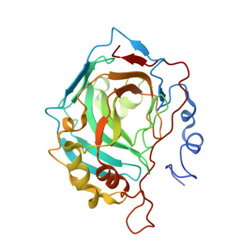
8R1I - PubMed Abstract:
The possible internal dynamics of non-isotope-labeled small-molecule ligands inside a target protein is inherently difficult to capture. Whereas high crystallographic temperature factors can denote either static disorder or motion, even moieties with very low B-factors can be subject to vivid motion between symmetry-related sites. Here we report the experimental identification of internal μs timescale dynamics of a high-affinity, natural-abundance ligand tightly bound to the enzyme human carbonic anhydrase II (hCAII) even within a crystalline lattice. The rotamer jumps of the ligand's benzene group manifest themselves both, in solution and fast magic-angle spinning solid-state NMR 1 H R 1ρ relaxation dispersion, for which we obtain further mechanistic insights from molecular-dynamics (MD) simulations. The experimental confirmation of rotameric jumps in bound ligands within proteins in solution or the crystalline state may improve understanding of host-guest interactions in biology and supra-molecular chemistry and may facilitate medicinal chemistry for future drug campaigns.
- Department of Chemistry and Chemical Biology, TU Dortmund University, Otto-Hahn-Str. 4a, 44227, Dortmund, Germany.
Organizational Affiliation: