8ROL

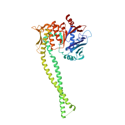
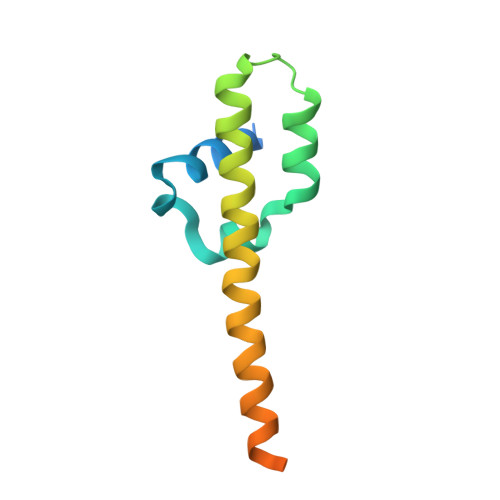
Human cohesin SMC3-HD(EQ)/RAD21-N complex - ATP-Mg-bound conformation - Form 2


Experimental Data Snapshot

  • Method: X-RAY DIFFRACTION
  • Resolution: 3.11 Å
  • R-Value Free: 0.238 
  • R-Value Work: 0.187 
  • R-Value Observed: 0.190 

Starting Model: experimental
View more details

wwPDB Validation   3D Report Full Report


Ligand Structure Quality Assessment 


This is version 1.2 of the entry. See complete history


Literature

The cohesin ATPase cycle is mediated by specific conformational dynamics and interface plasticity of SMC1A and SMC3 ATPase domains.

Vitoria Gomes, M.Landwerlin, P.Diebold-Durand, M.L.Shaik, T.B.Durand, A.Troesch, E.Weber, C.Brillet, K.Lemee, M.V.Decroos, C.Dulac, L.Antony, P.Watrin, E.Ennifar, E.Golzio, C.Romier, C.

(2024) Cell Rep 43: 114656-114656

  • DOI: https://doi.org/10.1016/j.celrep.2024.114656
  • Primary Citation of Related Structures:  
    8RO6, 8RO7, 8RO8, 8RO9, 8ROA, 8ROB, 8ROC, 8ROD, 8ROE, 8ROF, 8ROG, 8ROH, 8ROI, 8ROJ, 8ROK, 8ROL

  • PubMed Abstract: 

    Cohesin is key to eukaryotic genome organization and acts throughout the cell cycle in an ATP-dependent manner. The mechanisms underlying cohesin ATPase activity are poorly understood. Here, we characterize distinct steps of the human cohesin ATPase cycle and show that the SMC1A and SMC3 ATPase domains undergo specific but concerted structural rearrangements along this cycle. Specifically, whereas the proximal coiled coil of the SMC1A ATPase domain remains conformationally stable, that of the SMC3 displays an intrinsic flexibility. The ATP-dependent formation of the heterodimeric SMC1A/SMC3 ATPase module (engaged state) favors this flexibility, which is counteracted by NIPBL and DNA binding (clamped state). Opening of the SMC3/RAD21 interface (open-engaged state) stiffens the SMC3 proximal coiled coil, thus constricting together with that of SMC1A the ATPase module DNA-binding chamber. The plasticity of the ATP-dependent interface between the SMC1A and SMC3 ATPase domains enables these structural rearrangements while keeping the ATP gate shut. VIDEO ABSTRACT.


  • Organizational Affiliation

    Université de Strasbourg, IGBMC UMR 7104 - UMR-S 1258, 67400 Illkirch, France; CNRS, UMR 7104, 67400 Illkirch, France; INSERM, UMR-S 1258, 67400 Illkirch, France; Institut de Génétique et de Biologie Moléculaire et Cellulaire, Department of Integrated Structural Biology, 67400 Illkirch, France.


Macromolecules
Find similar proteins by:  (by identity cutoff)  |  3D Structure
Entity ID: 1
MoleculeChains Sequence LengthOrganismDetailsImage
Structural maintenance of chromosomes protein 3462Homo sapiensMutation(s): 1 
Gene Names: SMC3BAMBMHCSPG6SMC3L1
UniProt & NIH Common Fund Data Resources
Find proteins for Q9UQE7 (Homo sapiens)
Explore Q9UQE7 
Go to UniProtKB:  Q9UQE7
PHAROS:  Q9UQE7
GTEx:  ENSG00000108055 
Entity Groups  
Sequence Clusters30% Identity50% Identity70% Identity90% Identity95% Identity100% Identity
UniProt GroupQ9UQE7
Sequence Annotations
Expand
  • Reference Sequence
Find similar proteins by:  (by identity cutoff)  |  3D Structure
Entity ID: 2
MoleculeChains Sequence LengthOrganismDetailsImage
Double-strand-break repair protein rad21 homolog110Homo sapiensMutation(s): 0 
Gene Names: RAD21HR21KIAA0078NXP1SCC1
UniProt & NIH Common Fund Data Resources
Find proteins for O60216 (Homo sapiens)
Explore O60216 
Go to UniProtKB:  O60216
PHAROS:  O60216
GTEx:  ENSG00000164754 
Entity Groups  
Sequence Clusters30% Identity50% Identity70% Identity90% Identity95% Identity100% Identity
UniProt GroupO60216
Sequence Annotations
Expand
  • Reference Sequence
Small Molecules
Ligands 2 Unique
IDChains Name / Formula / InChI Key2D Diagram3D Interactions
AGS (Subject of Investigation/LOI)
Query on AGS

Download Ideal Coordinates CCD File 
D [auth A]PHOSPHOTHIOPHOSPHORIC ACID-ADENYLATE ESTER
C10 H16 N5 O12 P3 S
NLTUCYMLOPLUHL-KQYNXXCUSA-N
MG (Subject of Investigation/LOI)
Query on MG

Download Ideal Coordinates CCD File 
C [auth A]MAGNESIUM ION
Mg
JLVVSXFLKOJNIY-UHFFFAOYSA-N
Experimental Data & Validation

Experimental Data

  • Method: X-RAY DIFFRACTION
  • Resolution: 3.11 Å
  • R-Value Free: 0.238 
  • R-Value Work: 0.187 
  • R-Value Observed: 0.190 
  • Space Group: P 64 2 2
Unit Cell:
Length ( Å )Angle ( ˚ )
a = 201.067α = 90
b = 201.067β = 90
c = 92.376γ = 120
Software Package:
Software NamePurpose
PHENIXrefinement
XDSdata reduction
XDSdata scaling
PHENIXphasing

Structure Validation

View Full Validation Report


Ligand Structure Quality Assessment 


Entry History & Funding Information

Deposition Data


Funding OrganizationLocationGrant Number
Fondation ARCFranceARCPJA20181208268
Fondation ARCFranceARCPJA2021060003715
Fondation ARCFranceDOC20180507150
Agence Nationale de la Recherche (ANR)FranceANR-10-LABX-030-INRT
Agence Nationale de la Recherche (ANR)FranceANR-10-IDEX-0002
Agence Nationale de la Recherche (ANR)FranceANR-17-EURE-0023
Agence Nationale de la Recherche (ANR)FranceANR-10-INBS-0005-01

Revision History  (Full details and data files)

  • Version 1.0: 2024-09-11
    Type: Initial release
  • Version 1.1: 2024-09-18
    Changes: Database references
  • Version 1.2: 2024-09-25
    Changes: Structure summary