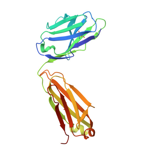
8TQ6 | pdb_00008tq6

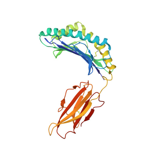
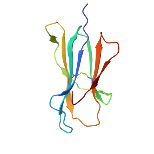
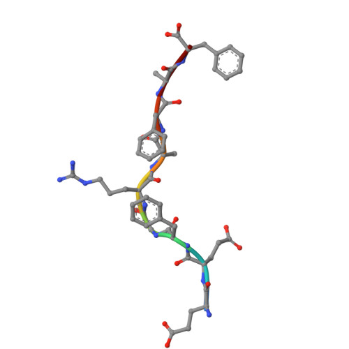
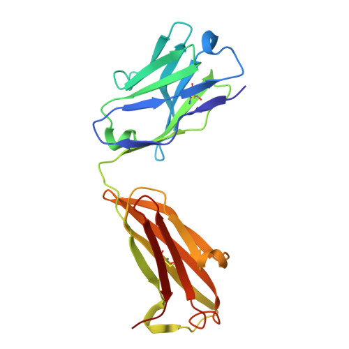
Crystal structure of Fab.B1.23.2 in complex with MHC-I (HLA-B*44:05)


Experimental Data Snapshot

  • Method: X-RAY DIFFRACTION
  • Resolution: 3.20 Å
  • R-Value Free: 
    0.268 (Depositor), 0.275 (DCC) 
  • R-Value Work: 
    0.240 (Depositor), 0.242 (DCC) 
  • R-Value Observed: 
    0.253 (Depositor) 

Starting Model: experimental
View more details

wwPDB Validation 3D Report Full Report

Validation slider image for 8TQ6

This is version 1.1 of the entry. See complete history. 

Literature

Structural mechanism of anti-MHC-I antibody blocking of inhibitory NK cell receptors in tumor immunity.

Jiang, J., Panda, A.K., Natarajan, K., Lei, H., Sharma, S., Boyd, L.F., Towler, R.R., Chempati, S., Ahmad, J., Morton, A.J., Lang, Z.C., Sun, Y., Sgourakis, N., Meier-Schellersheim, M., Huang, R.K., Shevach, E.M., Margulies, D.H.

(2026) Commun Biol 

  • DOI: https://doi.org/10.1038/s42003-026-09641-8
  • Primary Citation Related Structures: 
    8TQ6, 9D73, 9D74, 9OA9

  • PubMed Abstract: 

    Anti-major histocompatibility complex class I (MHC-I) mAbs can stimulate immune responses to tumors and infections by blocking suppressive signals delivered via various immune inhibitory receptors. To understand such functions, we determined the structure of a highly cross-reactive anti-human MHC-I mAb, B1.23.2, in complex with the MHC-I molecule HLA-B*44:05 by both cryo-electron microscopy (cryo-EM) and X-ray crystallography. Structural models determined by the two methods were essentially identical revealing that B1.23.2 binds a conserved region on the α2 1 helix that overlaps the killer immunoglobulin-like receptor (KIR) binding site. Structural comparison to KIR/HLA complexes reveals a mechanism by which B1.23.2 blocks inhibitory receptor interactions, leading to natural killer (NK) cell activation. B1.23.2 treatment of the human KLM-1 pancreatic cancer model in humanized (NSG-IL15) mice provides evidence of suppression of tumor growth. Such anti-MHC-I mAb that block inhibitory KIR/HLA interactions may prove useful for tumor immunotherapy.


  • Organizational Affiliation: 
    • Molecular Biology Section, Laboratory of Immune System Biology, National Institute of Allergy and Infectious Diseases, National Institutes of Health, Bethesda, MD, USA. jiangji@niaid.nih.gov.

Macromolecule Content 

  • Total Structure Weight: 182.3 kDa 
  • Atom Count: 12,516 
  • Modeled Residue Count: 1,607 
  • Deposited Residue Count: 1,616 
  • Unique protein chains: 5

Macromolecules

Find similar proteins by:|  3D Structure
Entity ID: 1
MoleculeChains  Sequence LengthOrganismDetailsImage
HLA class I histocompatibility antigen B alpha chain (HLA-B*44:05)
A, C
274Homo sapiensMutation(s): 0 
Gene Names: HLA-B
UniProt
Find proteins for Q860B7 (Homo sapiens)
Explore Q860B7 
Go to UniProtKB:  Q860B7
Entity Groups
Sequence Clusters30% Identity50% Identity70% Identity90% Identity95% Identity100% Identity
UniProt GroupQ860B7
Sequence Annotations
Expand
Reference Sequence
Find similar proteins by:|  3D Structure
Entity ID: 2
MoleculeChains  Sequence LengthOrganismDetailsImage
Beta-2-microglobulin
B, D
100Homo sapiensMutation(s): 0 
Gene Names: B2M, CDABP0092, HDCMA22P
UniProt & NIH Common Fund Data Resources
Find proteins for P61769 (Homo sapiens)
Explore P61769 
Go to UniProtKB:  P61769
PHAROS:  P61769
GTEx:  ENSG00000166710 
Entity Groups
Sequence Clusters30% Identity50% Identity70% Identity90% Identity95% Identity100% Identity
UniProt GroupP61769
Sequence Annotations
Expand
Reference Sequence
Find similar proteins by:  Sequence   |   3D Structure  
Entity ID: 3
MoleculeChains  Sequence LengthOrganismDetailsImage
MHC class II antigen peptideE,
J [auth P]
9synthetic constructMutation(s): 0 
UniProt
Find proteins for Q9TQB0 (Homo sapiens)
Explore Q9TQB0 
Go to UniProtKB:  Q9TQB0
Entity Groups
UniProt GroupQ9TQB0
Sequence Annotations
Expand
Reference Sequence
Find similar proteins by:|  3D Structure
Entity ID: 4
MoleculeChains  Sequence LengthOrganismDetailsImage
Fab B1.23.2 Heavy Chain
F, H
213Mus musculusMutation(s): 0 
Entity Groups
Sequence Clusters30% Identity50% Identity70% Identity90% Identity95% Identity100% Identity
Sequence Annotations
Expand
Reference Sequence
Find similar proteins by:|  3D Structure
Entity ID: 5
MoleculeChains  Sequence LengthOrganismDetailsImage
Fab B1.23.2 Light ChainG,
I [auth L]
212Mus musculusMutation(s): 0 
Entity Groups
Sequence Clusters30% Identity50% Identity70% Identity90% Identity95% Identity100% Identity
Sequence Annotations
Expand
Reference Sequence

Experimental Data & Validation

Experimental Data

  • Method: X-RAY DIFFRACTION
  • Resolution: 3.20 Å
  • R-Value Free:  0.268 (Depositor), 0.275 (DCC) 
  • R-Value Work:  0.240 (Depositor), 0.242 (DCC) 
  • R-Value Observed: 0.253 (Depositor) 
Space Group: P 21 21 21
Unit Cell:
Length ( Å )Angle ( ˚ )
a = 89.5α = 90
b = 92.84β = 90
c = 229.81γ = 90
Software Package:
Software NamePurpose
PHENIXrefinement
PHENIXrefinement
XDSdata reduction
XDSdata scaling
PHASERphasing

Structure Validation

View Full Validation Report


Entry History 

& Funding Information

Deposition Data


Funding OrganizationLocationGrant Number
National Institutes of Health/National Institute Of Allergy and Infectious Diseases (NIH/NIAID)United States--

Revision History  (Full details and data files)

  • Version 1.0: 2025-02-05
    Type: Initial release
  • Version 1.1: 2026-02-11
    Changes: Database references