8WQI | pdb_00008wqi

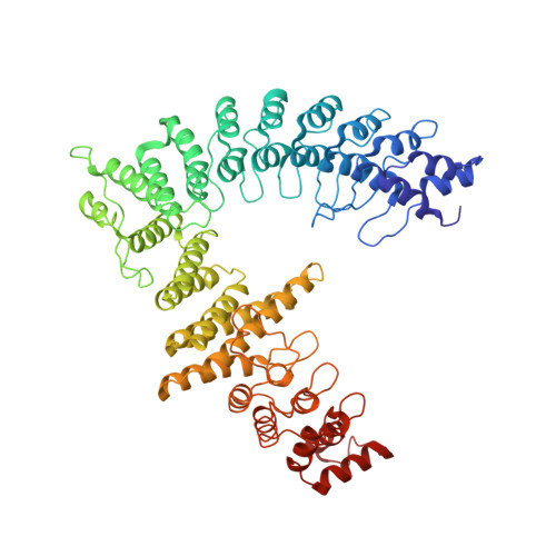
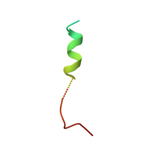
Local refinement of FEM1B bound with the C-degron of CUX1


Experimental Data Snapshot

  • Method: ELECTRON MICROSCOPY
  • Resolution: 3.50 Å
  • Aggregation State: PARTICLE 
  • Reconstruction Method: SINGLE PARTICLE 

wwPDB Validation 3D Report Full Report

Validation slider image for 8WQI

This is version 1.1 of the entry. See complete history. 

Literature

Mechanism of Psi-Pro/C-degron recognition by the CRL2 FEM1B ubiquitin ligase.

Chen, X., Raiff, A., Li, S., Guo, Q., Zhang, J., Zhou, H., Timms, R.T., Yao, X., Elledge, S.J., Koren, I., Zhang, K., Xu, C.

(2024) Nat Commun 15: 3558-3558

  • DOI: https://doi.org/10.1038/s41467-024-47890-5
  • Primary Citation Related Structures: 
    8WQA, 8WQB, 8WQC, 8WQD, 8WQE, 8WQF, 8WQG, 8WQH, 8WQI

  • PubMed Abstract: 

    The E3 ligase-degron interaction determines the specificity of the ubiquitin‒proteasome system. We recently discovered that FEM1B, a substrate receptor of Cullin 2-RING ligase (CRL2), recognizes C-degrons containing a C-terminal proline. By solving several cryo-EM structures of CRL2 FEM1B bound to different C-degrons, we elucidate the dimeric assembly of the complex. Furthermore, we reveal distinct dimerization states of unmodified and neddylated CRL2 FEM1B to uncover the NEDD8-mediated activation mechanism of CRL2 FEM1B . Our research also indicates that, FEM1B utilizes a bipartite mechanism to recognize both the C-terminal proline and an upstream aromatic residue within the substrate. These structural findings, complemented by in vitro ubiquitination and in vivo cell-based assays, demonstrate that CRL2 FEM1B -mediated polyubiquitination and subsequent protein turnover depend on both FEM1B-degron interactions and the dimerization state of the E3 ligase complex. Overall, this study deepens our molecular understanding of how Cullin-RING E3 ligase substrate selection mediates protein turnover.


  • Organizational Affiliation: 
    • MOE Key Laboratory for Cellular Dynamics, Division of Life Sciences and Medicine, University of Science and Technology of China, Hefei, 230027, PR China.

Macromolecule Content 

  • Total Structure Weight: 73.5 kDa 
  • Atom Count: 5,074 
  • Modeled Residue Count: 644 
  • Deposited Residue Count: 658 
  • Unique protein chains: 2

Macromolecules

Find similar proteins by:|  3D Structure
Entity ID: 1
MoleculeChains  Sequence LengthOrganismDetailsImage
Protein fem-1 homolog BA [auth D]627Homo sapiensMutation(s): 0 
Gene Names: FEM1B, F1AA, KIAA0396
UniProt & NIH Common Fund Data Resources
Find proteins for Q9UK73 (Homo sapiens)
Explore Q9UK73 
Go to UniProtKB:  Q9UK73
PHAROS:  Q9UK73
GTEx:  ENSG00000169018 
Entity Groups
Sequence Clusters30% Identity50% Identity70% Identity90% Identity95% Identity100% Identity
UniProt GroupQ9UK73
Sequence Annotations
Expand
Reference Sequence
Find similar proteins by:|  3D Structure
Entity ID: 2
MoleculeChains  Sequence LengthOrganismDetailsImage
Protein CASPB [auth G]31Homo sapiensMutation(s): 0 
Gene Names: CUX1, CUTL1
UniProt & NIH Common Fund Data Resources
Find proteins for Q13948 (Homo sapiens)
Explore Q13948 
Go to UniProtKB:  Q13948
PHAROS:  Q13948
GTEx:  ENSG00000257923 
Entity Groups
Sequence Clusters30% Identity50% Identity70% Identity90% Identity95% Identity100% Identity
UniProt GroupQ13948
Sequence Annotations
Expand
Reference Sequence

Experimental Data & Validation

Experimental Data

  • Method: ELECTRON MICROSCOPY
  • Resolution: 3.50 Å
  • Aggregation State: PARTICLE 
  • Reconstruction Method: SINGLE PARTICLE 
EM Software:
TaskSoftware PackageVersion
MODEL REFINEMENTPHENIX1.20.1_4487:

Structure Validation

View Full Validation Report


Entry History 

& Funding Information

Deposition Data


Funding OrganizationLocationGrant Number
National Natural Science Foundation of China (NSFC)China--

Revision History  (Full details and data files)

  • Version 1.0: 2024-04-03
    Type: Initial release
  • Version 1.1: 2024-05-08
    Changes: Database references