8ZC3

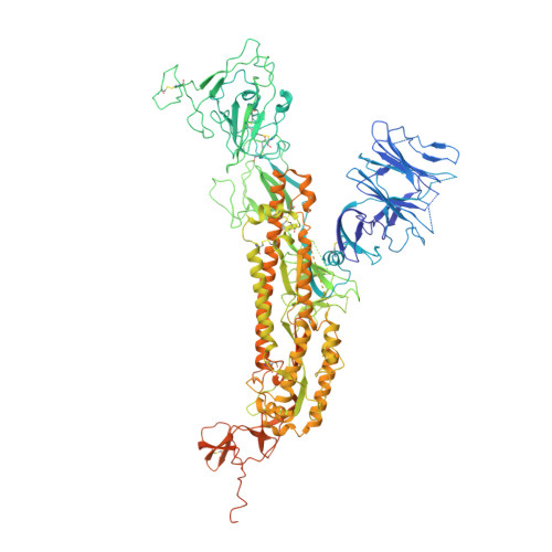
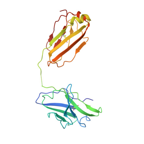
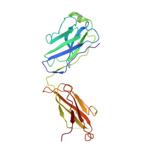
SARS-CoV-2 Omicron BA.4 spike trimer (6P) in complex with 3 D1F6 Fabs (1 RBD up)


Experimental Data Snapshot

  • Method: ELECTRON MICROSCOPY
  • Resolution: 4.69 Å
  • Aggregation State: PARTICLE 
  • Reconstruction Method: SINGLE PARTICLE 

wwPDB Validation   3D Report Full Report


This is version 1.3 of the entry. See complete history


Literature

An unconventional VH1-2 antibody tolerates escape mutations and shows an antigenic hotspot on SARS-CoV-2 spike.

Liu, B.Niu, X.Deng, Y.Zhang, Z.Wang, Y.Gao, X.Liang, H.Li, Z.Wang, Q.Cheng, Y.Chen, Q.Huang, S.Pan, Y.Su, M.Lin, X.Niu, C.Chen, Y.Yang, W.Zhang, Y.Yan, Q.He, J.Zhao, J.Chen, L.Xiong, X.

(2024) Cell Rep 43: 114265-114265

  • DOI: https://doi.org/10.1016/j.celrep.2024.114265
  • Primary Citation of Related Structures:  
    8ZBY, 8ZBZ, 8ZC0, 8ZC1, 8ZC2, 8ZC3, 8ZC4, 8ZC5, 8ZC6

  • PubMed Abstract: 

    The severe acute respiratory syndrome coronavirus 2 (SARS-CoV-2) spike (S) protein continues to evolve antigenically, impacting antibody immunity. D1F6, an affinity-matured non-stereotypic VH1-2 antibody isolated from a patient infected with the SARS-CoV-2 ancestral strain, effectively neutralizes most Omicron variants tested, including XBB.1.5. We identify that D1F6 in the immunoglobulin G (IgG) form is able to overcome the effect of most Omicron mutations through its avidity-enhanced multivalent S-trimer binding. Cryo-electron microscopy (cryo-EM) and biochemical analyses show that three simultaneous epitope mutations are generally needed to substantially disrupt the multivalent S-trimer binding by D1F6 IgG. Antigenic mutations at spike positions 346, 444, and 445, which appeared in the latest variants, have little effect on D1F6 binding individually. However, these mutations are able to act synergistically with earlier Omicron mutations to impair neutralization by affecting the interaction between D1F6 IgG and the S-trimer. These results provide insight into the mechanism by which accumulated antigenic mutations facilitate evasion of affinity-matured antibodies.


  • Organizational Affiliation

    State Key Laboratory of Respiratory Disease, CAS Key Laboratory of Regenerative Biology, Guangdong Provincial Key Laboratory of Stem Cell and Regenerative Medicine, Guangdong Provincial Key Laboratory of Biocomputing, Guangzhou Institutes of Biomedicine and Health, Chinese Academy of Sciences, Guangzhou, China.


Macromolecules
Find similar proteins by:  (by identity cutoff)  |  3D Structure
Entity ID: 1
MoleculeChains Sequence LengthOrganismDetailsImage
Spike glycoprotein
A, B, C
1,238Severe acute respiratory syndrome coronavirus 2Mutation(s): 34 
Gene Names: S2
UniProt
Find proteins for P0DTC2 (Severe acute respiratory syndrome coronavirus 2)
Explore P0DTC2 
Go to UniProtKB:  P0DTC2
Entity Groups  
Sequence Clusters30% Identity50% Identity70% Identity90% Identity95% Identity100% Identity
UniProt GroupP0DTC2
Glycosylation
Glycosylation Sites: 13Go to GlyGen: P0DTC2-1
Sequence Annotations
Expand
  • Reference Sequence
Find similar proteins by:  (by identity cutoff)  |  3D Structure
Entity ID: 2
MoleculeChains Sequence LengthOrganismDetailsImage
Light chain of D1F6 FabD,
F [auth M],
G [auth N]
223Homo sapiensMutation(s): 0 
Entity Groups  
Sequence Clusters30% Identity50% Identity70% Identity90% Identity95% Identity100% Identity
Sequence Annotations
Expand
  • Reference Sequence
Find similar proteins by:  (by identity cutoff)  |  3D Structure
Entity ID: 3
MoleculeChains Sequence LengthOrganismDetailsImage
Heavy chain of D1F6 FabE,
H [auth Q],
I [auth R]
230Homo sapiensMutation(s): 0 
Entity Groups  
Sequence Clusters30% Identity50% Identity70% Identity90% Identity95% Identity100% Identity
Sequence Annotations
Expand
  • Reference Sequence
Oligosaccharides

Help

Entity ID: 4
MoleculeChains Length2D Diagram Glycosylation3D Interactions
2-acetamido-2-deoxy-beta-D-glucopyranose-(1-4)-2-acetamido-2-deoxy-beta-D-glucopyranose
J [auth F],
K [auth G],
L [auth H],
M [auth I],
N [auth J],
J [auth F],
K [auth G],
L [auth H],
M [auth I],
N [auth J],
O [auth K],
P [auth L],
Q [auth O],
R [auth P],
S,
T
2N-Glycosylation
Glycosylation Resources
GlyTouCan:  G42666HT
GlyCosmos:  G42666HT
GlyGen:  G42666HT
Small Molecules
Ligands 1 Unique
IDChains Name / Formula / InChI Key2D Diagram3D Interactions
NAG (Subject of Investigation/LOI)
Query on NAG

Download Ideal Coordinates CCD File 
AA [auth A]
BA [auth A]
CA [auth A]
DA [auth A]
EA [auth B]
AA [auth A],
BA [auth A],
CA [auth A],
DA [auth A],
EA [auth B],
FA [auth B],
GA [auth B],
HA [auth B],
IA [auth B],
JA [auth B],
KA [auth B],
LA [auth B],
MA [auth B],
NA [auth C],
OA [auth C],
PA [auth C],
QA [auth C],
RA [auth C],
SA [auth C],
TA [auth C],
U [auth A],
UA [auth C],
V [auth A],
VA [auth C],
W [auth A],
X [auth A],
Y [auth A],
Z [auth A]
2-acetamido-2-deoxy-beta-D-glucopyranose
C8 H15 N O6
OVRNDRQMDRJTHS-FMDGEEDCSA-N
Experimental Data & Validation

Experimental Data

  • Method: ELECTRON MICROSCOPY
  • Resolution: 4.69 Å
  • Aggregation State: PARTICLE 
  • Reconstruction Method: SINGLE PARTICLE 

Structure Validation

View Full Validation Report


Entry History & Funding Information

Deposition Data


Funding OrganizationLocationGrant Number
Other governmentEKPG21-06
Other governmentSRPG22-002
Other government2021A1515011289

Revision History  (Full details and data files)

  • Version 1.0: 2024-05-15
    Type: Initial release
  • Version 1.1: 2024-05-22
    Changes: Structure summary
  • Version 1.2: 2024-06-12
    Changes: Database references
  • Version 1.3: 2024-11-06
    Changes: Data collection, Structure summary