Download Mendeley
Cryo-EM and cryo-ET reveal the molecular architecture and host interactions of mycobacteriophage Douge.
Maharana, J., Wang, C.H., Tsai, L.A., Liao, Y.T., Yang, C.H., Shen, M.C., Macale, L.S., Tran, T.N., Narsico, J., Perez, R.J., Tewary, S.K., Wu, J.L., Lin, H.Y., Chang, S.W., Franklin, A., Moynihan, P.J., Jacobs-Sera, D., Freeman, K.G., Hatfull, G.F., Lowary, T.L., Ho, M.C.(2025) Cell Rep 44: 116057-116057
- PubMed: 40711877 Search on PubMedSearch on PubMed Central
- DOI: https://doi.org/10.1016/j.celrep.2025.116057
- Primary Citation Related Structures:
8ZDH, 8ZDI, 8ZDJ, 8ZDK, 8ZDL, 8ZDM, 8ZDN, 8ZDO, 8ZDP, 8ZDQ, 8ZEA - PubMed Abstract:
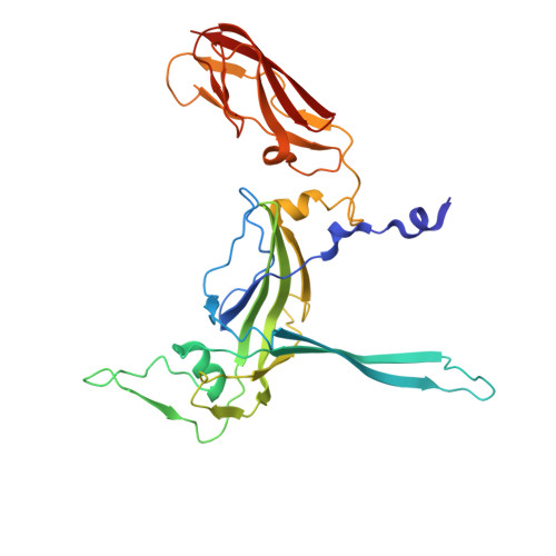
Recent reports highlight the efficacy of engineered mycobacteriophages to treat non-tuberculosis mycobacterial disease. Molecular insights into mycobacteriophage architecture and host interactions could allow structure-guided phage engineering to increase efficacy and broaden host range, but such information is currently unavailable. We describe the cryoelectron microscopy (cryo-EM) structure of mycobacteriophage Douge, which contains 1,105 protein subunits assembled into a complete siphophage and is coated with glycan-binding domains for mycobacterial cell surface interactions. When filled with viral genome, the channel spanning the connector, tail, and baseplate is sealed by tape measure proteins, providing a genome gating system and requiring limited structural changes for genome ejection upon phage-host contact. Nanometer-resolution cryoelectron tomography (cryo-ET) snapshots of phage-host interactions show that the baseplate remains attached to the mycobacterial outer membrane during viral genome ejection. This study reveals high-resolution structural details of this mycobacteriophage and its interaction with host glycans.
- Institute of Biological Chemistry, Academia Sinica, Taipei, Taiwan; Taiwan International Graduate Program in Chemical Biology and Molecular Biophysics (TIGP-CBMB), Academia Sinica, Taipei, Taiwan; Institute of Bioinformatics and Structural Biology, College of Life Sciences and Medicine, National Tsing Hua University, Hsinchu, Taiwan.
Organizational Affiliation: