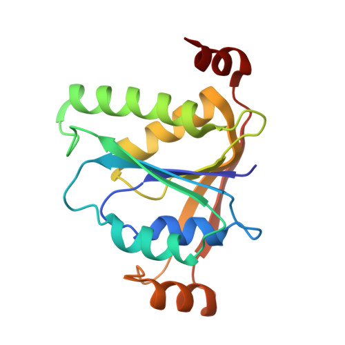
9C6A | pdb_00009c6a

The CRISPR associated adenosine deaminase Cad1-CARF in the apo form


Experimental Data Snapshot

  • Method: X-RAY DIFFRACTION
  • Resolution: 3.60 Å
  • R-Value Free: 
    0.317 (Depositor), 0.317 (DCC) 
  • R-Value Work: 
    0.248 (Depositor), 0.248 (DCC) 
  • R-Value Observed: 
    0.252 (Depositor) 

Starting Model: in silico
View more details

wwPDB Validation 3D Report Full Report

Validation slider image for 9C6A

This is version 1.2 of the entry. See complete history. 

Literature

The CRISPR-associated adenosine deaminase Cad1 converts ATP to ITP to provide antiviral immunity.

Baca, C.F., Majumder, P., Hickling, J.H., Ye, L., Teplova, M., Brady, S.F., Patel, D.J., Marraffini, L.A.

(2024) Cell 187: 7183

  • DOI: https://doi.org/10.1016/j.cell.2024.10.002
  • Primary Citation Related Structures: 
    9C67, 9C68, 9C69, 9C6A, 9C6C, 9C6F, 9C77, 9CDB

  • PubMed Abstract: 

    Type III CRISPR systems provide immunity against genetic invaders through the production of cyclic oligo-adenylate (cA n ) molecules that activate effector proteins that contain CRISPR-associated Rossman fold (CARF) domains. Here, we characterized the function and structure of an effector in which the CARF domain is fused to an adenosine deaminase domain, CRISPR-associated adenosine deaminase 1 (Cad1). We show that upon binding of cA 4 or cA 6 to its CARF domain, Cad1 converts ATP to ITP, both in vivo and in vitro. Cryoelectron microscopy (cryo-EM) structural studies on full-length Cad1 reveal an hexameric assembly composed of a trimer of dimers, with bound ATP at inter-domain sites required for activity and ATP/ITP within deaminase active sites. Upon synthesis of cA n during phage infection, Cad1 activation leads to a growth arrest of the host that prevents viral propagation. Our findings reveal that CRISPR-Cas systems employ a wide range of molecular mechanisms beyond nucleic acid degradation to provide adaptive immunity in prokaryotes.


  • Organizational Affiliation: 
    • Laboratory of Bacteriology, The Rockefeller University, New York, NY 10065, USA; Tri-Institutional PhD Program in Chemical Biology, Weill Cornell Medical College, Rockefeller University and Memorial Sloan Kettering Cancer Center, New York, NY 10065, USA.

Macromolecule Content 

  • Total Structure Weight: 75.86 kDa 
  • Atom Count: 5,340 
  • Modeled Residue Count: 663 
  • Deposited Residue Count: 664 
  • Unique protein chains: 1

Macromolecules

Find similar proteins by:|  3D Structure
Entity ID: 1
MoleculeChains  Sequence LengthOrganismDetailsImage
Adenosine deaminase domain-containing protein
A, B, C, D
166Bacteroidales bacteriumMutation(s): 0 
Gene Names: DCM62_02910
Entity Groups
Sequence Clusters30% Identity50% Identity70% Identity90% Identity95% Identity100% Identity
Sequence Annotations
Expand
Reference Sequence

Experimental Data & Validation

Experimental Data

  • Method: X-RAY DIFFRACTION
  • Resolution: 3.60 Å
  • R-Value Free:  0.317 (Depositor), 0.317 (DCC) 
  • R-Value Work:  0.248 (Depositor), 0.248 (DCC) 
  • R-Value Observed: 0.252 (Depositor) 
Space Group: P 21 21 21
Unit Cell:
Length ( Å )Angle ( ˚ )
a = 53.615α = 90
b = 96.801β = 90
c = 155.722γ = 90
Software Package:
Software NamePurpose
PHENIXrefinement
Aimlessdata scaling
MOLREPphasing
HKL-2000data reduction

Structure Validation

View Full Validation Report


Entry History 

& Funding Information

Deposition Data


Funding OrganizationLocationGrant Number
National Institutes of Health/National Institute of General Medical Sciences (NIH/NIGMS)United StatesNIHGM129430, NIHGM145888

Revision History  (Full details and data files)

  • Version 1.0: 2024-10-30
    Type: Initial release
  • Version 1.1: 2024-11-06
    Changes: Database references
  • Version 1.2: 2024-12-25
    Changes: Database references