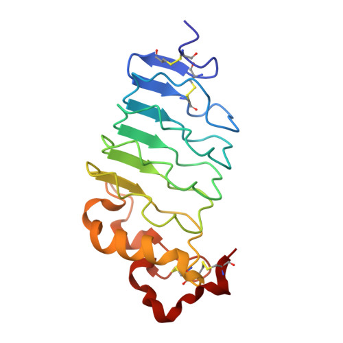
9CJ0

The High-Resolution Structure of a Variable Lymphocyte Receptor from Petromyzon marinus Capable of Binding to the Brain Extracellular Matrix


Experimental Data Snapshot

  • Method: X-RAY DIFFRACTION
  • Resolution: 1.30 Å
  • R-Value Free: 0.222 
  • R-Value Work: 0.185 
  • R-Value Observed: 0.186 

Starting Model: experimental
View more details

wwPDB Validation   3D Report Full Report


Ligand Structure Quality Assessment 


This is version 1.3 of the entry. See complete history


Literature

The High-Resolution Structure of a Variable Lymphocyte Receptor From Petromyzon marinus Capable of Binding to the Brain Extracellular Matrix.

Appelt, E.A.Thoden, J.B.Gehrke, S.A.Bachmeier, H.D.Rayment, I.Shusta, E.V.Holden, H.M.

(2024) Proteins 

  • DOI: https://doi.org/10.1002/prot.26768
  • Primary Citation of Related Structures:  
    9CJ0

  • PubMed Abstract: 

    Variable lymphocyte receptors (VLRs) are antigen receptors derived from the adaptive immune system of jawless vertebrates such as lamprey (Petromyzon marinus). First discovered in 2004, VLRs have been the subject of numerous biochemical and structural investigations. Due to their unique antigen binding properties, VLRs have been leveraged as possible drug delivery agents. One such VLR, previously identified and referred to as P1C10, was shown to bind to the brain extracellular matrix. Here, we present the high-resolution X-ray crystal structure of this VLR determined to 1.3 Å resolution. The fold is dominated by a six-stranded mixed β-sheet which provides a concave surface for possible antigen binding. Electron density corresponding to a 4-(2-hydroxyethyl)piperazine-1-propanesulfonic acid buffer molecule (HEPPS) was found in this region. By comparing the P1C10 molecular architecture and its buffer binding residues with those of other VLRs previously reported, it was possible to illustrate how this unique class of proteins can accommodate diverse binding partners. Additionally, we provide an analysis of the experimentally determined structure compared to the models generated by the commonly used AlphaFold and iTASSER structure prediction software packages.


  • Organizational Affiliation

    Department of Chemical and Biological Engineering, University of Wisconsin-Madison, Madison, Wisconsin, USA.


Macromolecules
Find similar proteins by:  (by identity cutoff)  |  3D Structure
Entity ID: 1
MoleculeChains Sequence LengthOrganismDetailsImage
Variable Lymphocyte Receptor170Petromyzon marinusMutation(s): 0 
Entity Groups  
Sequence Clusters30% Identity50% Identity70% Identity90% Identity95% Identity100% Identity
Sequence Annotations
Expand
  • Reference Sequence
Experimental Data & Validation

Experimental Data

  • Method: X-RAY DIFFRACTION
  • Resolution: 1.30 Å
  • R-Value Free: 0.222 
  • R-Value Work: 0.185 
  • R-Value Observed: 0.186 
  • Space Group: P 21 21 21
Unit Cell:
Length ( Å )Angle ( ˚ )
a = 45.522α = 90
b = 53.137β = 90
c = 60.348γ = 90
Software Package:
Software NamePurpose
REFMACrefinement
SAINTdata reduction
SADABSdata scaling
PHASERphasing

Structure Validation

View Full Validation Report


Ligand Structure Quality Assessment 


Entry History & Funding Information

Deposition Data


Funding OrganizationLocationGrant Number
National Institutes of Health/National Institute of General Medical Sciences (NIH/NIGMS)United StatesR35 GM134643
National Institutes of Health/National Institute of General Medical Sciences (NIH/NIGMS)United States5 T32 GM135066
National Institutes of Health/National Institute of Neurological Disorders and Stroke (NIH/NINDS)United StatesR01 NS099158
National Institutes of Health/National Institute of Neurological Disorders and Stroke (NIH/NINDS)United StatesR01 NS118028

Revision History  (Full details and data files)

  • Version 1.0: 2024-07-31
    Type: Initial release
  • Version 1.1: 2024-09-04
    Changes: Database references, Structure summary
  • Version 1.2: 2024-10-23
    Changes: Structure summary
  • Version 1.3: 2024-12-11
    Changes: Database references