Structures and functions of the limited natural polyclonal antibody response to parvovirus infection.
Adu, O.F., Lee, H., Fruh, S.P., Schoenle, M.V., Weichert, W.S., Flyak, A.I., Hafenstein, S.L., Parrish, C.R.(2025) Proc Natl Acad Sci U S A 122: e2423460122-e2423460122
- PubMed: 39951487 Search on PubMed
- DOI: https://doi.org/10.1073/pnas.2423460122
- Primary Citation Related Structures:
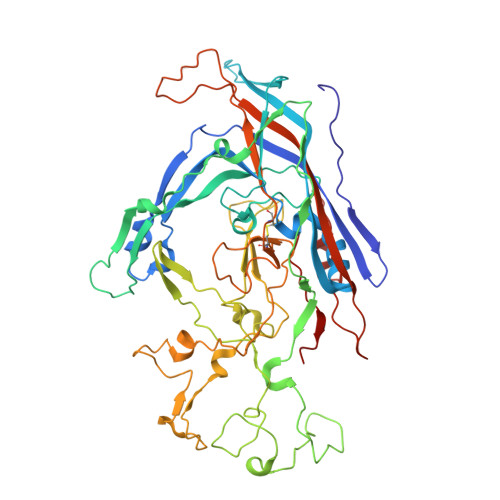
9E60, 9E7W, 9E7X, 9E7Z, 9E80, 9E89, 9E8D - PubMed Abstract:
Host antibody responses are key components in the protection of animals against pathogens, yet the defining properties of viral antigens and induction of B cell responses that result in varied protection are still poorly understood. Parvoviruses are simple molecular structures that display 60 repeated motifs on their capsid surface, and rapidly induce strong antibody responses that protect animals from infection. We recently showed that following canine parvovirus infection of its natural host, the polyclonal response in the sera contained only two or three dominant antibodies that bound two epitopes on the capsid. Here, we characterize key antibodies present in that immune response, identifying their sequences, defining their binding properties on the capsid by cryoelectron microscopic (cryoEM) analysis, and testing their effects on viral infectivity. Two antibodies sharing the same heavy chain bound to the side of the capsid threefold spike (B-site), while another distinct antibody bound close to the threefold axis (A-site). The epitopes of these antibodies overlapped the binding site of the host receptor, the transferrin receptor type-1, but to varying degrees. The antibodies varied widely in their neutralization efficiencies as either immunoglobulins (IgGs) or monomeric antigen-binding fragments (Fabs), which was consistent with their ability to compete for the receptor. The monoclonal antibodies characterized here matched the structures from the cryoEM analysis of polyclonal sera, including those present in a different dog than the monoclonal source. This shows that after infection, a focused response to the viral antigen is produced that protects against infection.
- Department of Microbiology and Immunology, College of Veterinary Medicine, Cornell University, Ithaca, NY 14853.
Organizational Affiliation: