


Biochemical and structural insights into the auto-inhibited state of Mical1 and its activation by Rab8
Rai, A., Janning, P., Vetter, I.R., Goody, R.S.(2024) Elife
Experimental Data Snapshot
Starting Model: experimental
View more details
(2024) Elife
Entity ID: 1 | |||||
|---|---|---|---|---|---|
| Molecule | Chains | Sequence Length | Organism | Details | Image |
| [F-actin]-monooxygenase MICAL1 | A [auth B] | 153 | Homo sapiens | Mutation(s): 1 Gene Names: MICAL1, MICAL, NICAL EC: 1.14.13.225 (PDB Primary Data), 1.6.3.1 (PDB Primary Data) | 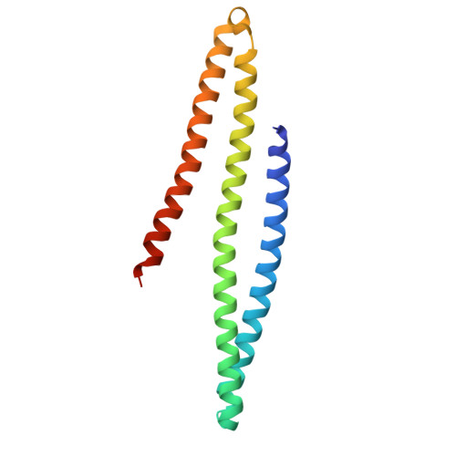 |
UniProt & NIH Common Fund Data Resources | |||||
PHAROS: Q8TDZ2 GTEx: ENSG00000135596 | |||||
Entity Groups | |||||
| Sequence Clusters | 30% Identity50% Identity70% Identity90% Identity95% Identity100% Identity | ||||
| UniProt Group | Q8TDZ2 | ||||
Sequence AnnotationsExpand | |||||
Reference Sequence | |||||
Entity ID: 2 | |||||
|---|---|---|---|---|---|
| Molecule | Chains | Sequence Length | Organism | Details | Image |
| Ras-related protein Rab-10 | B [auth C] | 177 | Homo sapiens | Mutation(s): 0 Gene Names: RAB10 EC: 3.6.5.2 | 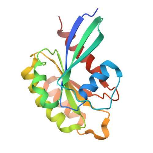 |
UniProt & NIH Common Fund Data Resources | |||||
PHAROS: P61026 GTEx: ENSG00000084733 | |||||
Entity Groups | |||||
| Sequence Clusters | 30% Identity50% Identity70% Identity90% Identity95% Identity100% Identity | ||||
| UniProt Group | P61026 | ||||
Sequence AnnotationsExpand | |||||
Reference Sequence | |||||
| Ligands 2 Unique | |||||
|---|---|---|---|---|---|
| ID | Chains | Name / Formula / InChI Key | 2D Diagram | 3D Interactions | |
| GNP (Subject of Investigation/LOI) Download:Ideal Coordinates CCD File | C | PHOSPHOAMINOPHOSPHONIC ACID-GUANYLATE ESTER C10 H17 N6 O13 P3 UQABYHGXWYXDTK-UUOKFMHZSA-N |  | ||
| MG (Subject of Investigation/LOI) Download:Ideal Coordinates CCD File | D [auth C] | MAGNESIUM ION Mg JLVVSXFLKOJNIY-UHFFFAOYSA-N |  | ||
| Length ( Å ) | Angle ( ˚ ) |
|---|---|
| a = 53.77 | α = 90 |
| b = 49.65 | β = 98.54 |
| c = 79.33 | γ = 90 |
| Software Name | Purpose |
|---|---|
| PHENIX | refinement |
| XDS | data reduction |
| XSCALE | data scaling |
| PHASER | phasing |
| Funding Organization | Location | Grant Number |
|---|---|---|
| Max Planck Society | Germany | -- |
| German Research Foundation (DFG) | Germany | grant GO 284/10-1 |


