9M3P | pdb_00009m3p

Crystal structure of human pyruvate dehydrogenase kinase isoform 1 in complex with ATP competitive inhibitor 3


Experimental Data Snapshot

  • Method: X-RAY DIFFRACTION
  • Resolution: 2.01 Å
  • R-Value Free: 
    0.196 (Depositor), 0.199 (DCC) 
  • R-Value Work: 
    0.163 (Depositor), 0.166 (DCC) 
  • R-Value Observed: 
    0.165 (Depositor) 

Starting Model: experimental
View more details

wwPDB Validation 3D Report Full Report

Validation slider image for 9M3P

Ligand Structure Quality Assessment 


This is version 1.0 of the entry. See complete history

Literature

Discovery of ATP competitive PDHK1/2 dual inhibitors.

Xu, H.Ding, D.Han, X.Miao, K.Liang, C.Yun, H.Zhu, W.Dey, F.Zhao, D.Wu, Y.Reutlinger, M.Yang, J.Zhai, G.Lin, Z.Li, C.Wu, W.Xu, B.Han, L.Chen, S.Huang, X.Casagrande, F.Hilbert, M.Strebel, Q.Wichert, M.Westwood, P.Schafer, R.Roth, D.Heer, D.Tian, X.Ma, T.Zhang, T.Zhao, J.Urich, E.Xia, G.Lassen, K.Shen, H.C.Zou, G.

(2025) Bioorg Med Chem Lett 122: 130190-130190

  • DOI: https://doi.org/10.1016/j.bmcl.2025.130190
  • Primary Citation Related Structures: 
    9M3O, 9M3P, 9M3R, 9M3T, 9M3U

  • PubMed Abstract: 

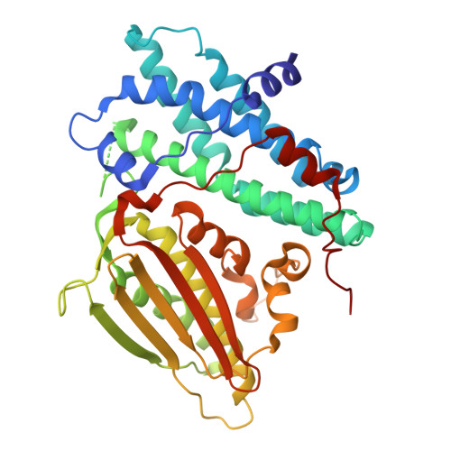
    Multiple screening approaches were carried out to identify novel chemistry starting for Pyruvate Dehydrogenase Kinases (PDHKs) inhibitors. Through hit triaging efforts and structure-based optimization, two series of ATP competitive inhibitors with single digit nanomolar enzymatic potency for PDHK1/2 and around 10-100-fold selectivity over PDHK4/3 were discovered. Approach of covalent inhibitor was explored to successfully improve the cellular target engagement to single digit micromolar range.


  • Organizational Affiliation
    • China Innovation Center of Roche, No. 371 Lishizhen Road, Shanghai, 201203, China.

Macromolecule Content 

  • Total Structure Weight: 45.21 kDa 
  • Atom Count: 3,391 
  • Modeled Residue Count: 373 
  • Deposited Residue Count: 383 
  • Unique protein chains: 1

Macromolecules

Find similar proteins by:|  3D Structure
Entity ID: 1
MoleculeChains  Sequence LengthOrganismDetailsImage
[Pyruvate dehydrogenase (acetyl-transferring)] kinase isozyme 1, mitochondrial383Homo sapiensMutation(s): 0 
Gene Names: PDK1PDHK1
EC: 2.7.11.2
UniProt & NIH Common Fund Data Resources
Find proteins for Q15118 (Homo sapiens)
Explore Q15118 
Go to UniProtKB:  Q15118
PHAROS:  Q15118
GTEx:  ENSG00000152256 
Entity Groups
Sequence Clusters30% Identity50% Identity70% Identity90% Identity95% Identity100% Identity
UniProt GroupQ15118
Sequence Annotations
Expand
Reference Sequence

Small Molecules

Ligands 3 Unique
IDChains Name / Formula / InChI Key2D Diagram3D Interactions
SZ6
(Subject of Investigation/LOI)

Query on SZ6


Download:Ideal Coordinates CCD File
C [auth A],
D [auth A]
N-[4-(2-chloro-5-methylpyrimidin-4-yl)phenyl]-N-(4-{[(difluoroacetyl)amino]methyl}benzyl)-2,4-dihydroxybenzamide
C28 H23 Cl F2 N4 O4
LCGNLQSOSJFLKR-UHFFFAOYSA-N
GOL

Query on GOL


Download:Ideal Coordinates CCD File
E [auth A],
F [auth A],
G [auth A]
GLYCEROL
C3 H8 O3
PEDCQBHIVMGVHV-UHFFFAOYSA-N
K

Query on K


Download:Ideal Coordinates CCD File
B [auth A]POTASSIUM ION
K
NPYPAHLBTDXSSS-UHFFFAOYSA-N

Experimental Data & Validation

Experimental Data

  • Method: X-RAY DIFFRACTION
  • Resolution: 2.01 Å
  • R-Value Free:  0.196 (Depositor), 0.199 (DCC) 
  • R-Value Work:  0.163 (Depositor), 0.166 (DCC) 
  • R-Value Observed: 0.165 (Depositor) 
Space Group: P 41 21 2
Unit Cell:
Length ( Å )Angle ( ˚ )
a = 100.221α = 90
b = 100.221β = 90
c = 109.815γ = 90
Software Package:
Software NamePurpose
PHENIXrefinement
XDSdata reduction
Aimlessdata scaling
PHASERphasing

Structure Validation

View Full Validation Report


Ligand Structure Quality Assessment 


Entry History 

& Funding Information

Deposition Data


Funding OrganizationLocationGrant Number
Not funded--

Revision History  (Full details and data files)

  • Version 1.0: 2025-04-02
    Type: Initial release