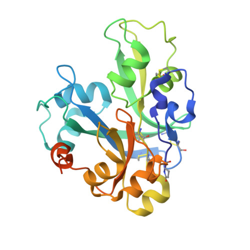
9Q84 | pdb_00009q84

Crystal structure of Lotus japonicus CHIP13 extracellular domain in complex with chitooctaose


Experimental Data Snapshot

  • Method: X-RAY DIFFRACTION
  • Resolution: 1.41 Å
  • R-Value Free: 
    0.181 (Depositor), 0.181 (DCC) 
  • R-Value Work: 
    0.146 (Depositor), 0.146 (DCC) 
  • R-Value Observed: 
    0.148 (Depositor) 

Starting Model: experimental
View more details

wwPDB Validation 3D Report Full Report

Validation slider image for 9Q84

This is version 1.2 of the entry. See complete history. 

Literature

Structural basis for size-selective perception of chitin in plants.

Gysel, K., Hansen, S.B., Rubsam, H., Alsarraf, H.M.A.B., Madland, E., Baadegaard, C., Engholm, E., Cheng, J.X.J., Poulsen, E.C., Vinther, M., Jensen, K.J., Thygesen, M.B., Fort, S., Stougaard, J., Andersen, K.R.

(2026) Science 394: 298-305

  • DOI: https://doi.org/10.1126/science.adz4600
  • Primary Citation Related Structures: 
    9H6V, 9Q83, 9Q84, 9QRS

  • PubMed Abstract: 

    Plants detect microbes through pattern recognition receptors that perceive conserved microbial surface motifs known as microbe-associated molecular patterns (MAMPs). Lysin motif (LysM) receptors recognize and mediate downstream responses to chitinous MAMPs. In this study, we elucidated the mechanism for the specific recognition of long-chain chitin oligomers and identified a hallmark bridge domain characteristic of the CHITIN PERCEPTION (CHIP) receptor class. Structural analysis of receptor-ligand complexes, biochemistry, and in planta functional studies of inhibitory nanobodies revealed the mechanism of size-selective, high-affinity chitin perception in Lotus japonicus and Medicago truncatula . Additionally, we identified the CHITIN ELICITOR RECEPTOR KINASE (CERK) class as low-affinity, yet essential, co-receptors and propose a mechanistic model for a ligand-induced core signaling heterocomplex. Our findings establish the structural and mechanistic basis of differential chitin perception and receptor complex assembly in chitin-elicited plant immune signaling.


  • Organizational Affiliation: 
    • Department of Molecular Biology and Genetics, Aarhus University, Aarhus, Denmark.

Macromolecule Content 

  • Total Structure Weight: 31.69 kDa 
  • Atom Count: 2,279 
  • Modeled Residue Count: 222 
  • Deposited Residue Count: 248 
  • Unique protein chains: 1

Macromolecules

Find similar proteins by:|  3D Structure
Entity ID: 1
MoleculeChains  Sequence LengthOrganismDetailsImage
LysM type receptor kinase248Lotus japonicusMutation(s): 0 
Gene Names: LYS13
UniProt
Find proteins for D3KU00 (Lotus japonicus)
Explore D3KU00 
Go to UniProtKB:  D3KU00
Entity Groups
Sequence Clusters30% Identity50% Identity70% Identity90% Identity95% Identity100% Identity
UniProt GroupD3KU00
Glycosylation
Glycosylation Sites: 4
Sequence Annotations
Expand
Reference Sequence

Oligosaccharides

Help  
Entity ID: 2
MoleculeChains Length2D Diagram GlycosylationD Interactions
2-acetamido-2-deoxy-beta-D-glucopyranose-(1-4)-2-acetamido-2-deoxy-beta-D-glucopyranose-(1-4)-2-acetamido-2-deoxy-beta-D-glucopyranose-(1-4)-2-acetamido-2-deoxy-beta-D-glucopyranose-(1-4)-2-acetamido-2-deoxy-beta-D-glucopyranose-(1-4)-2-acetamido-2-deoxy-beta-D-glucopyranose-(1-4)-2-acetamido-2-deoxy-beta-D-glucopyranose-(1-4)-2-acetamido-2-deoxy-beta-D-glucopyranose
B
8N/A
Glycosylation Resources
GlyTouCan: G83257UC
GlyCosmos: G83257UC
Entity ID: 3
MoleculeChains Length2D Diagram GlycosylationD Interactions
2-acetamido-2-deoxy-beta-D-glucopyranose-(1-4)-2-acetamido-2-deoxy-beta-D-glucopyranose
C, D
2N-Glycosylation
Glycosylation Resources
GlyTouCan: G42666HT
GlyCosmos: G42666HT
GlyGen: G42666HT
Entity ID: 4
MoleculeChains Length2D Diagram GlycosylationD Interactions
beta-D-mannopyranose-(1-4)-2-acetamido-2-deoxy-beta-D-glucopyranose-(1-4)-2-acetamido-2-deoxy-beta-D-glucopyranose
E
3N-Glycosylation
Glycosylation Resources
GlyTouCan: G15407YE
GlyCosmos: G15407YE
GlyGen: G15407YE
Entity ID: 5
MoleculeChains Length2D Diagram GlycosylationD Interactions
alpha-D-mannopyranose-(1-3)-[alpha-D-mannopyranose-(1-6)]beta-D-mannopyranose-(1-4)-2-acetamido-2-deoxy-beta-D-glucopyranose-(1-4)-[alpha-L-fucopyranose-(1-6)]2-acetamido-2-deoxy-beta-D-glucopyranose
F
6N-Glycosylation
Glycosylation Resources
GlyTouCan: G82348BZ
GlyCosmos: G82348BZ
GlyGen: G82348BZ

Small Molecules

Modified Residues  1 Unique
IDChains Modified ResidueModification TypeFormula2D Diagram
PCA
Query on PCA
GLNPyrrolidone carboxylic acidC5 H7 N O3

Experimental Data & Validation

Experimental Data

  • Method: X-RAY DIFFRACTION
  • Resolution: 1.41 Å
  • R-Value Free:  0.181 (Depositor), 0.181 (DCC) 
  • R-Value Work:  0.146 (Depositor), 0.146 (DCC) 
  • R-Value Observed: 0.148 (Depositor) 
Space Group: C 1 2 1
Unit Cell:
Length ( Å )Angle ( ˚ )
a = 73.882α = 90
b = 66.776β = 109.348
c = 53.203γ = 90
Software Package:
Software NamePurpose
PHENIXrefinement
XDSdata reduction
XSCALEdata scaling
PHASERphasing

Structure Validation

View Full Validation Report


Entry History 

& Funding Information

Deposition Data


Funding OrganizationLocationGrant Number
The Carlsberg FoundationDenmarkCF21-0139
Danish Council for Independent ResearchDenmark3103-00137B
Novo Nordisk FoundationDenmarkNNF21OC0071300

Revision History  (Full details and data files)

  • Version 1.0: 2025-07-16
    Type: Initial release
  • Version 1.1: 2026-10-07
    Changes: Database references
  • Version 1.2: 2026-10-28
    Changes: Database references