2G8N | pdb_00002g8n

Structure of hPNMT with inhibitor 3-Hydroxymethyl-7-(N-4-chlorophenylaminosulfonyl)-THIQ and AdoHcy


Experimental Data Snapshot

  • Method: X-RAY DIFFRACTION
  • Resolution: 2.15 Å
  • R-Value Free: 
    0.243 (Depositor), 0.239 (DCC) 
  • R-Value Work: 
    0.201 (Depositor), 0.198 (DCC) 

Starting Model: experimental
View more details

wwPDB Validation   3D Report Full Report


Ligand Structure Quality Assessment 

Created with Raphaël 2.3.0Worse 01 BetterLigand structure goodness of fit to experimental dataBest fitted SAHClick on this verticalbar to view detailsBest fitted F83Click on this verticalbar to view details

This is version 1.4 of the entry. See complete history


Literature

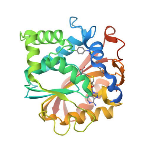
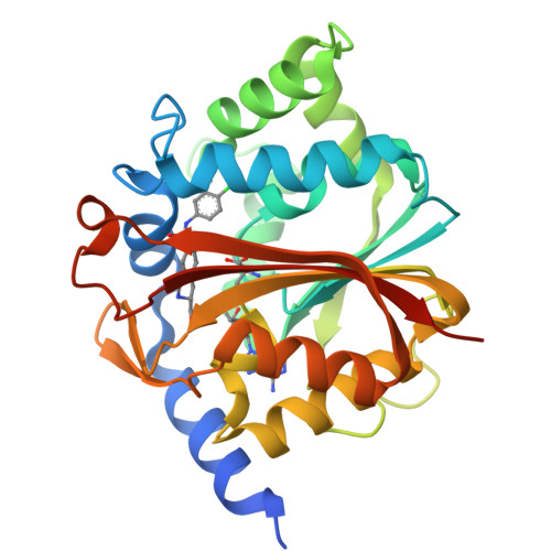
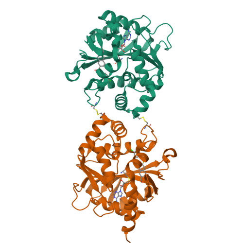
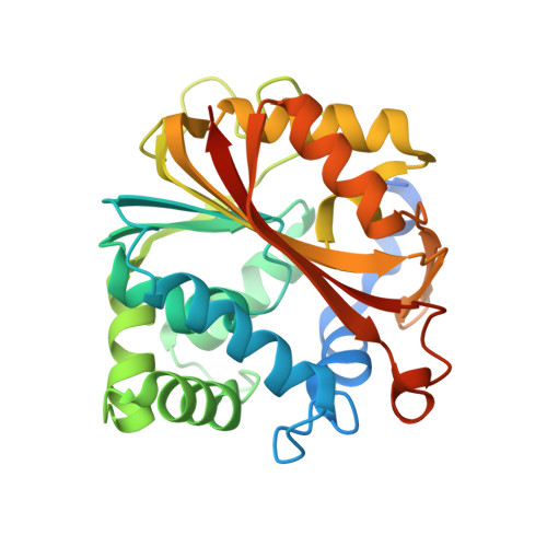
Comparison of the Binding of 3-Fluoromethyl-7-sulfonyl-1,2,3,4-tetrahydroisoquinolines with Their Isosteric Sulfonamides to the Active Site of Phenylethanolamine N-Methyltransferase

Grunewald, G.L.Seim, M.R.Regier, R.C.Martin, J.L.Gee, C.L.Drinkwater, N.Criscione, K.R.

(2006) J Med Chem 49: 5424-5433

  • DOI: https://doi.org/10.1021/jm060466d
  • Primary Citation of Related Structures:  
    2G8N

  • PubMed Abstract: 

    3-Fluoromethyl-7-(N-substituted aminosulfonyl)-1,2,3,4-tetrahydroisoquinolines (14, 16, and 18-22) are highly potent and selective inhibitors of phenylethanolamine N-methyltransferase (PNMT). Molecular modeling studies with 3-fluoromethyl-7-(N-alkyl aminosulfonyl)-1,2,3,4-tetrahydroisoquinolines, such as 16, suggested that the sulfonamide -NH- could form a hydrogen bond with the side chain of Lys57. However, SAR studies and analysis of the crystal structure of human PNMT (hPNMT) in complex with 7 indicated that the sulfonamide oxygens, and not the sulfonamide -NH-, formed favorable interactions with the enzyme. Thus, we hypothesized that replacement of the sulfonamide -NH- with a methylene group could result in compounds that would retain potency at PNMT and that would have increased lipophilicity, thus increasing the likelihood they will cross the blood brain barrier. A series of 3-fluoromethyl-7-sulfonyl-1,2,3,4-tetrahydroisoquinolines (23-30) were synthesized and evaluated for their PNMT inhibitory potency and affinity for the alpha2-adrenoceptor. A comparison of these compounds with their isosteric sulfonamides (14, 16, and 18-22) showed that the sulfones were more lipophilic but less potent than their corresponding sulfonamides. Sulfone 24 (hPNMT Ki = 1.3 microM) is the most potent compound in this series and is quite selective for PNMT versus the alpha2-adrenoceptor, but 24 is less potent than the corresponding sulfonamide, 16 (hPNMT Ki = 0.13 microM). We also report the crystal structure of hPNMT in complex with sulfonamide 15, from which a potential hydrogen bond acceptor within the hPNMT active site has been identified, the main chain carbonyl oxygen of Asn39. The interaction of this residue with the sulfonamide -NH- is likely responsible for much of the enhanced inhibitory potency of the sulfonamides versus the sulfones.


  • Organizational Affiliation

    Department of Medicinal Chemistry, University of Kansas, Lawrence, Kansas 66045, USA. ggrunewald@ku.edu


Macromolecules
Find similar proteins by:  (by identity cutoff)  |  3D Structure
Entity ID: 1
MoleculeChains Sequence LengthOrganismDetailsImage
Phenylethanolamine N-methyltransferase
A, B
289Homo sapiensMutation(s): 0 
EC: 2.1.1.28
UniProt & NIH Common Fund Data Resources
Find proteins for P11086 (Homo sapiens)
Explore P11086 
Go to UniProtKB:  P11086
PHAROS:  P11086
GTEx:  ENSG00000141744 
Entity Groups  
Sequence Clusters30% Identity50% Identity70% Identity90% Identity95% Identity100% Identity
UniProt GroupP11086
Sequence Annotations
Expand
  • Reference Sequence
Small Molecules
Binding Affinity Annotations 
IDSourceBinding Affinity
F83 BindingDB:  2G8N Ki: min: 39, max: 63 (nM) from 2 assay(s)
SAH BindingDB:  2G8N Ki: 1.40e+4 (nM) from 1 assay(s)
Experimental Data & Validation

Experimental Data

  • Method: X-RAY DIFFRACTION
  • Resolution: 2.15 Å
  • R-Value Free:  0.243 (Depositor), 0.239 (DCC) 
  • R-Value Work:  0.201 (Depositor), 0.198 (DCC) 
Space Group: P 43 21 2
Unit Cell:
Length ( Å )Angle ( ˚ )
a = 94.32α = 90
b = 94.32β = 90
c = 188.88γ = 90
Software Package:
Software NamePurpose
d*TREKdata processing
CNSrefinement
PDB_EXTRACTdata extraction
CrystalCleardata reduction
d*TREKdata scaling
CNSphasing

Structure Validation

View Full Validation Report


Ligand Structure Quality Assessment 

Created with Raphaël 2.3.0Worse 01 BetterLigand structure goodness of fit to experimental dataBest fitted SAHClick on this verticalbar to view detailsBest fitted F83Click on this verticalbar to view details

Entry History 

Deposition Data

Revision History  (Full details and data files)

  • Version 1.0: 2006-09-12
    Type: Initial release
  • Version 1.1: 2008-05-01
    Changes: Version format compliance
  • Version 1.2: 2011-07-13
    Changes: Derived calculations, Version format compliance
  • Version 1.3: 2023-10-25
    Changes: Data collection, Database references, Derived calculations, Refinement description
  • Version 1.4: 2024-10-23
    Changes: Data collection, Structure summary