4ZTE | pdb_00004zte

Crystal structure of human E-Cadherin (residues 3-213) in complex with a peptidomimetic inhibitor


Experimental Data Snapshot

  • Method: X-RAY DIFFRACTION
  • Resolution: 2.13 Å
  • R-Value Free: 
    0.230 (Depositor), 0.230 (DCC) 
  • R-Value Work: 
    0.185 (Depositor), 0.190 (DCC) 
  • R-Value Observed: 
    0.187 (Depositor) 

Starting Model: experimental
View more details

wwPDB Validation   3D Report Full Report


Ligand Structure Quality Assessment 

Created with Raphaël 2.3.0Worse 01 BetterLigand structure goodness of fit to experimental dataBest fitted 4RLClick on this verticalbar to view details

This is version 1.2 of the entry. See complete history


Literature

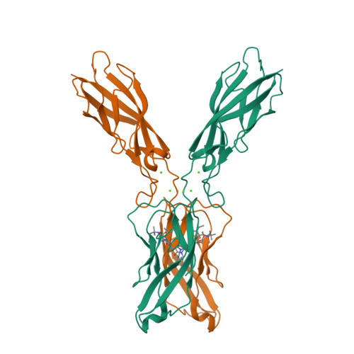
Crystal Structure of Human E-Cadherin-EC1EC2 in Complex with a Peptidomimetic Competitive Inhibitor of Cadherin Homophilic Interaction.

Nardone, V.Lucarelli, A.P.Dalle Vedove, A.Fanelli, R.Tomassetti, A.Belvisi, L.Civera, M.Parisini, E.

(2016) J Med Chem 59: 5089-5094

  • DOI: https://doi.org/10.1021/acs.jmedchem.5b01487
  • Primary Citation of Related Structures:  
    4ZT1, 4ZTE

  • PubMed Abstract: 

    Cadherins are transmembrane cell adhesion proteins whose aberrant expression often correlates with cancer development and proliferation. We report the crystal structure of an E-cadherin extracellular fragment in complex with a peptidomimetic compound that was previously shown to partially inhibit cadherin homophilic adhesion. The structure reveals an unexpected binding mode and allows the identification of a druggable cadherin interface, thus paving the way to a future structure-guided design of cell adhesion inhibitors against cadherin-expressing solid tumors.


  • Organizational Affiliation

    Center for Nano Science and Technology @PoliMi, Istituto Italiano di Tecnologia , Via G. Pascoli 70/3, 20133 Milano, Italy.


Macromolecules
Find similar proteins by:  (by identity cutoff)  |  3D Structure
Entity ID: 1
MoleculeChains Sequence LengthOrganismDetailsImage
Cadherin-1
A, B
211Homo sapiensMutation(s): 0 
Gene Names: CDH1CDHEUVO
UniProt & NIH Common Fund Data Resources
Find proteins for P12830 (Homo sapiens)
Explore P12830 
Go to UniProtKB:  P12830
PHAROS:  P12830
GTEx:  ENSG00000039068 
Entity Groups  
Sequence Clusters30% Identity50% Identity70% Identity90% Identity95% Identity100% Identity
UniProt GroupP12830
Sequence Annotations
Expand
  • Reference Sequence
Small Molecules
Ligands 2 Unique
IDChains Name / Formula / InChI Key2D Diagram3D Interactions
4RL
Query on 4RL

Download Ideal Coordinates CCD File 
F [auth A],
J [auth B]
N-{[(2S,5S)-1-benzyl-5-(2-{[(2S,3S)-1-(tert-butylamino)-3-methyl-1-oxopentan-2-yl]amino}-2-oxoethyl)-3,6-dioxopiperazin-2-yl]methyl}-L-alpha-asparagine
C28 H42 N6 O7
QOSIRQNYWJNZFB-WGLQCKHLSA-N
CA
Query on CA

Download Ideal Coordinates CCD File 
C [auth A]
D [auth A]
E [auth A]
G [auth B]
H [auth B]
CALCIUM ION
Ca
BHPQYMZQTOCNFJ-UHFFFAOYSA-N
Experimental Data & Validation

Experimental Data

  • Method: X-RAY DIFFRACTION
  • Resolution: 2.13 Å
  • R-Value Free:  0.230 (Depositor), 0.230 (DCC) 
  • R-Value Work:  0.185 (Depositor), 0.190 (DCC) 
  • R-Value Observed: 0.187 (Depositor) 
Space Group: C 1 2 1
Unit Cell:
Length ( Å )Angle ( ˚ )
a = 120.579α = 90
b = 76.835β = 111.14
c = 72.772γ = 90
Software Package:
Software NamePurpose
REFMACrefinement
SCALAdata scaling
MOLREPphasing
XDSdata reduction

Structure Validation

View Full Validation Report


Ligand Structure Quality Assessment 

Created with Raphaël 2.3.0Worse 01 BetterLigand structure goodness of fit to experimental dataBest fitted 4RLClick on this verticalbar to view details

Entry History & Funding Information

Deposition Data


Funding OrganizationLocationGrant Number
European UnionFP7-PEOPLE-2010-RG (N. 268231)

Revision History  (Full details and data files)

  • Version 1.0: 2016-06-01
    Type: Initial release
  • Version 1.1: 2016-06-08
    Changes: Database references
  • Version 1.2: 2024-01-10
    Changes: Data collection, Database references, Derived calculations, Refinement description