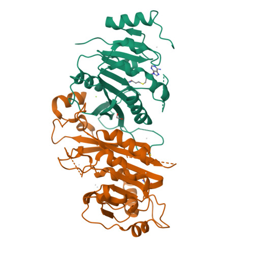
Human METTL3-METTL14 complex
ZENG, H., DONG, A., LI, Y., TEMPEL, W., Bountra, C., Arrowsmith, C.H., Edwards, A.M., BROWN, P.J., WU, H., Structural Genomics Consortium (SGC)To be published.
Experimental Data Snapshot
Starting Model: experimental
View more details
Entity ID: 1 | |||||
|---|---|---|---|---|---|
| Molecule | Chains | Sequence Length | Organism | Details | Image |
| N6-adenosine-methyltransferase 70 kDa subunit | 598 | Homo sapiens | Mutation(s): 0 Gene Names: METTL3, MTA70 EC: 2.1.1.62 (PDB Primary Data), 2.1.1.348 (UniProt) |  | |
UniProt & NIH Common Fund Data Resources | |||||
Find proteins for Q86U44 (Homo sapiens) Explore Q86U44 Go to UniProtKB: Q86U44 | |||||
PHAROS: Q86U44 GTEx: ENSG00000165819 | |||||
Entity Groups | |||||
| Sequence Clusters | 30% Identity50% Identity70% Identity90% Identity95% Identity100% Identity | ||||
| UniProt Group | Q86U44 | ||||
Sequence AnnotationsExpand | |||||
| |||||
Entity ID: 2 | |||||
|---|---|---|---|---|---|
| Molecule | Chains | Sequence Length | Organism | Details | Image |
| N6-adenosine-methyltransferase subunit METTL14 | 399 | Homo sapiens | Mutation(s): 0 Gene Names: METTL14, KIAA1627 EC: 2.1.1.62 |  | |
UniProt & NIH Common Fund Data Resources | |||||
Find proteins for Q9HCE5 (Homo sapiens) Explore Q9HCE5 Go to UniProtKB: Q9HCE5 | |||||
PHAROS: Q9HCE5 GTEx: ENSG00000145388 | |||||
Entity Groups | |||||
| Sequence Clusters | 30% Identity50% Identity70% Identity90% Identity95% Identity100% Identity | ||||
| UniProt Group | Q9HCE5 | ||||
Sequence AnnotationsExpand | |||||
| |||||
| Ligands 5 Unique | |||||
|---|---|---|---|---|---|
| ID | Chains | Name / Formula / InChI Key | 2D Diagram | 3D Interactions | |
| SAH Query on SAH | C [auth A] | S-ADENOSYL-L-HOMOCYSTEINE C14 H20 N6 O5 S ZJUKTBDSGOFHSH-WFMPWKQPSA-N |  | ||
| BME Query on BME | H [auth B] | BETA-MERCAPTOETHANOL C2 H6 O S DGVVWUTYPXICAM-UHFFFAOYSA-N |  | ||
| EDO Query on EDO | E [auth A] | 1,2-ETHANEDIOL C2 H6 O2 LYCAIKOWRPUZTN-UHFFFAOYSA-N |  | ||
| MG Query on MG | D [auth A] | MAGNESIUM ION Mg JLVVSXFLKOJNIY-UHFFFAOYSA-N |  | ||
| UNX Query on UNX | F [auth A] G [auth A] I [auth B] J [auth B] K [auth B] | UNKNOWN ATOM OR ION X |  | ||
| Length ( Å ) | Angle ( ˚ ) |
|---|---|
| a = 63.883 | α = 90 |
| b = 63.883 | β = 90 |
| c = 225.709 | γ = 120 |
| Software Name | Purpose |
|---|---|
| SCALEPACK | data scaling |
| REFMAC | refinement |
| PDB_EXTRACT | data extraction |
| PHASER | phasing |
| HKL-3000 | data reduction |
RCSB PDB is hosted by

RCSB PDB is a member of the