12KO | pdb_000012ko

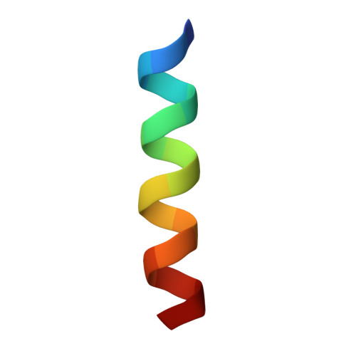
Supramolecular assembly of a staphylococcal delta-toxin peptide fragment, STIP3-1


Experimental Data Snapshot

  • Method: X-RAY DIFFRACTION
  • Resolution: 1.70 Å
  • R-Value Free: 
    0.243 (Depositor), 0.243 (DCC) 
  • R-Value Work: 
    0.197 (Depositor), 0.197 (DCC) 
  • R-Value Observed: 
    0.201 (Depositor) 

Starting Model: experimental
View more details

wwPDB Validation 3D Report Full Report

Validation slider image for 12KO

Ligand Structure Quality Assessment 


This is version 1.0 of the entry. See complete history. 

Literature

Supramolecular Assembly of an Antimicrobial Peptide Into a Pore.

Deeyagahage, K., Dhindwal, P., Boniecki, M.T., Ruzzini, A.

(2026) Angew Chem Int Ed Engl : e2822225-e2822225

  • DOI: https://doi.org/10.1002/anie.2822225
  • Primary Citation Related Structures: 
    12KM, 12KN, 12KO

  • PubMed Abstract: 

    Antimicrobial peptides (AMPs) have long been ascribed the ability to form pores. These structures, some observed, others inferred, vary in their mechanisms of action, though most are associated with lysis. Here, we show that an AMP derived from a truncated staphylococcal δ-toxin can form functional pores that do not cause rapid lysis but, instead, we suggest that they act as self-transport systems. Specifically, a 17-amino acid AMP named STIP3-1 was observed to oligomerize and form a unique supramolecular helical assembly with an ∼14 Å pore. Substitutions to the STIP3-1 peptide abolished our ability to detect pore formation in model membranes and prevented peptide accumulation in Staphylococcus aureus. In the case of G3A and G3Aib variants, the substitutions resulted in stabilization of antiparallel 4-helix bundles rather than a nanotubular assembly characterized by a two-ring structure formed by repeating trimers. MD simulations suggest that Gly may impart versatility in STIP3-1 phospholipid binding. Our observation of this remarkable STIP3-1 structure expands how we can approach mechanistic and translational studies of AMPs.


  • Organizational Affiliation: 
    • Department of Veterinary Microbiology, University of Saskatchewan, Saskatoon, Saskatchewan, Canada.

Macromolecule Content 

  • Total Structure Weight: 5.92 kDa 
  • Atom Count: 410 
  • Modeled Residue Count: 49 
  • Deposited Residue Count: 51 
  • Unique protein chains: 1

Macromolecules

Find similar proteins by:  Sequence   |   3D Structure  
Entity ID: 1
MoleculeChains  Sequence LengthOrganismDetailsImage
THR-VAL-GLY-GLY-LEU-VAL-LYS-TRP-ILE-LEU-ASP-THR-VAL-LYS-LYS-PHE-ALAA [auth B],
B [auth A],
C
17Staphylococcus auricularisMutation(s): 0 
Entity Groups
Sequence Clusters30% Identity50% Identity70% Identity90% Identity95% Identity100% Identity
Sequence Annotations
Expand
Reference Sequence

Experimental Data & Validation

Experimental Data

  • Method: X-RAY DIFFRACTION
  • Resolution: 1.70 Å
  • R-Value Free:  0.243 (Depositor), 0.243 (DCC) 
  • R-Value Work:  0.197 (Depositor), 0.197 (DCC) 
  • R-Value Observed: 0.201 (Depositor) 
Space Group: P 65
Unit Cell:
Length ( Å )Angle ( ˚ )
a = 49.748α = 90
b = 49.748β = 90
c = 30.1γ = 120
Software Package:
Software NamePurpose
PHENIXrefinement
XSCALEdata scaling
XDSdata reduction
PHASERphasing
PDB_EXTRACTdata extraction

Structure Validation

View Full Validation Report


Ligand Structure Quality Assessment 


Entry History 

& Funding Information

Deposition Data


Funding OrganizationLocationGrant Number
Natural Sciences and Engineering Research Council (NSERC, Canada)Canada--
Other governmentCanadaPOC.16.21
Other governmentCanada2022F142

Revision History  (Full details and data files)

  • Version 1.0: 2026-10-14
    Type: Initial release