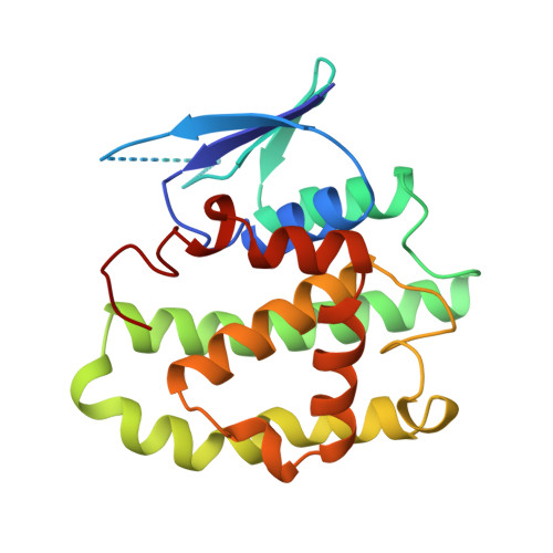
14GS

GLUTATHIONE S-TRANSFERASE P1-1 APO FORM 1


Experimental Data Snapshot

  • Method: X-RAY DIFFRACTION
  • Resolution: 2.80 Å
  • R-Value Free: 0.240 
  • R-Value Work: 0.190 
  • R-Value Observed: 0.190 

wwPDB Validation   3D Report Full Report


This is version 1.3 of the entry. See complete history


Literature

Evidence for an induced-fit mechanism operating in pi class glutathione transferases.

Oakley, A.J.Lo Bello, M.Ricci, G.Federici, G.Parker, M.W.

(1998) Biochemistry 37: 9912-9917

  • DOI: https://doi.org/10.1021/bi980323w
  • Primary Citation of Related Structures:  
    14GS, 16GS

  • PubMed Abstract: 

    Three-dimensional structures of the apo form of human pi class glutathione transferase have been determined by X-ray crystallography. The structures suggest the enzyme recognizes its substrate, glutathione, by an induced-fit mechanism. Compared to complexed forms of the enzyme, the environment around the catalytic residue, Tyr 7, remains unchanged in the apoenzyme. This observation supports the view that Tyr 7 does not act as a general base in the reaction mechanism. The observed cooperativity of the dimeric enzyme may be due to the movements of a helix that forms one wall of the active site and, in particular, to movements of a tyrosine residue that is located in the subunit interface.


  • Organizational Affiliation

    The Ian Potter Foundation Protein Crystallography Laboratory, St. Vincent's Institute of Medical Research, Fitzroy, Victoria, Australia.


Macromolecules
Find similar proteins by:  (by identity cutoff)  |  3D Structure
Entity ID: 1
MoleculeChains Sequence LengthOrganismDetailsImage
GLUTATHIONE S-TRANSFERASE
A, B
210Homo sapiensMutation(s): 0 
Gene Names: GSTP1
EC: 2.5.1.18
UniProt & NIH Common Fund Data Resources
Find proteins for P09211 (Homo sapiens)
Explore P09211 
Go to UniProtKB:  P09211
PHAROS:  P09211
GTEx:  ENSG00000084207 
Entity Groups  
Sequence Clusters30% Identity50% Identity70% Identity90% Identity95% Identity100% Identity
UniProt GroupP09211
Sequence Annotations
Expand
  • Reference Sequence
Small Molecules
Ligands 1 Unique
IDChains Name / Formula / InChI Key2D Diagram3D Interactions
MES
Query on MES

Download Ideal Coordinates CCD File 
C [auth A],
D [auth B]
2-(N-MORPHOLINO)-ETHANESULFONIC ACID
C6 H13 N O4 S
SXGZJKUKBWWHRA-UHFFFAOYSA-N
Experimental Data & Validation

Experimental Data

  • Method: X-RAY DIFFRACTION
  • Resolution: 2.80 Å
  • R-Value Free: 0.240 
  • R-Value Work: 0.190 
  • R-Value Observed: 0.190 
  • Space Group: C 1 2 1
Unit Cell:
Length ( Å )Angle ( ˚ )
a = 78.651α = 90
b = 91.826β = 98.25
c = 69.143γ = 90
Software Package:
Software NamePurpose
AMoREphasing
X-PLORrefinement
DENZOdata reduction
SCALEPACKdata scaling

Structure Validation

View Full Validation Report


Entry History 

Deposition Data

Revision History  (Full details and data files)

  • Version 1.0: 1999-01-13
    Type: Initial release
  • Version 1.1: 2008-03-03
    Changes: Version format compliance
  • Version 1.2: 2011-07-13
    Changes: Version format compliance
  • Version 1.3: 2023-08-02
    Changes: Database references, Derived calculations, Refinement description