RCSB PDB - 1A02: STRUCTURE OF THE DNA BINDING DOMAINS OF NFAT, FOS AND JUN BOUND TO DNA

 1A02

STRUCTURE OF THE DNA BINDING DOMAINS OF NFAT, FOS AND JUN BOUND TO DNA


Experimental Data Snapshot

  • Method: X-RAY DIFFRACTION
  • Resolution: 2.70 Å
  • R-Value Free: 0.303 
  • R-Value Work: 0.246 
  • R-Value Observed: 0.246 

wwPDB Validation   3D Report Full Report


This is version 1.4 of the entry. See complete history


Literature

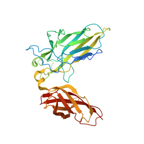
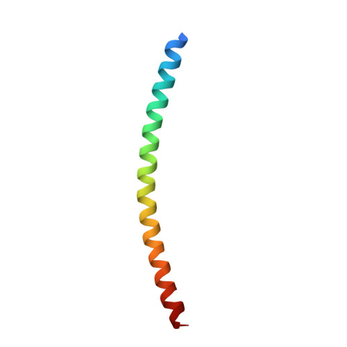
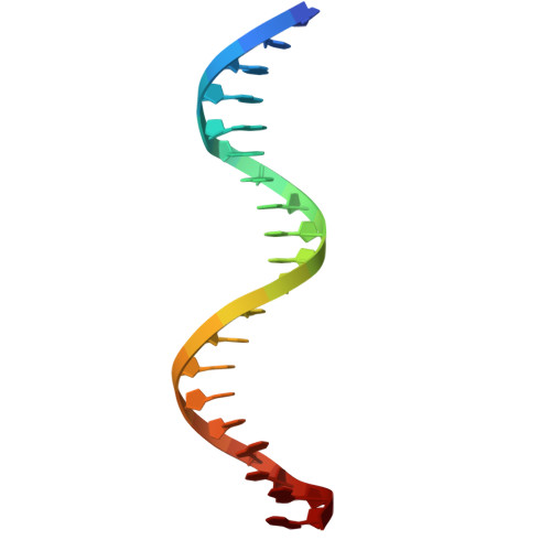
Structure of the DNA-binding domains from NFAT, Fos and Jun bound specifically to DNA.

Chen, L.Glover, J.N.Hogan, P.G.Rao, A.Harrison, S.C.

(1998) Nature 392: 42-48

  • DOI: https://doi.org/10.1038/32100
  • Primary Citation of Related Structures:  
    1A02

  • PubMed Abstract: 

    The nuclear factor of activated T cells (NFAT) and the AP-1 heterodimer, Fos-Jun, cooperatively bind a composite DNA site and synergistically activate the expression of many immune-response genes. A 2.7-A-resolution crystal structure of the DNA-binding domains of NFAT, Fos and Jun, in a quaternary complex with a DNA fragment containing the distal antigen-receptor response element from the interleukin-2 gene promoter, shows an extended interface between NFAT and AP-1, facilitated by the bending of Fos and DNA. The tight association of the three proteins on DNA creates a continuous groove for the recognition of 15 base pairs.


  • Organizational Affiliation

    Department of Molecular and Cellular Biology, Harvard University, Cambridge, Massachusetts 02138, USA. lchen@xta1200.harvard.edu


Macromolecules

Find similar proteins by:  (by identity cutoff)  |  3D Structure
Entity ID: 3
MoleculeChains Sequence LengthOrganismDetailsImage
NUCLEAR FACTOR OF ACTIVATED T CELLSC [auth N]301Homo sapiensMutation(s): 0 
Gene Names: NFAT1
UniProt & NIH Common Fund Data Resources
Find proteins for Q13469 (Homo sapiens)
Explore Q13469 
Go to UniProtKB:  Q13469
PHAROS:  Q13469
GTEx:  ENSG00000101096 
Entity Groups  
Sequence Clusters30% Identity50% Identity70% Identity90% Identity95% Identity100% Identity
UniProt GroupQ13469
Sequence Annotations
Expand
  • Reference Sequence
Find similar proteins by:  (by identity cutoff)  |  3D Structure
Entity ID: 4
MoleculeChains Sequence LengthOrganismDetailsImage
AP-1 FRAGMENT FOSD [auth F]56Homo sapiensMutation(s): 2 
UniProt & NIH Common Fund Data Resources
Find proteins for P01100 (Homo sapiens)
Explore P01100 
Go to UniProtKB:  P01100
PHAROS:  P01100
GTEx:  ENSG00000170345 
Entity Groups  
Sequence Clusters30% Identity50% Identity70% Identity90% Identity95% Identity100% Identity
UniProt GroupP01100
Sequence Annotations
Expand
  • Reference Sequence
Find similar proteins by:  (by identity cutoff)  |  3D Structure
Entity ID: 5
MoleculeChains Sequence LengthOrganismDetailsImage
AP-1 FRAGMENT JUNE [auth J]56Homo sapiensMutation(s): 2 
UniProt & NIH Common Fund Data Resources
Find proteins for P05412 (Homo sapiens)
Explore P05412 
Go to UniProtKB:  P05412
PHAROS:  P05412
GTEx:  ENSG00000177606 
Entity Groups  
Sequence Clusters30% Identity50% Identity70% Identity90% Identity95% Identity100% Identity
UniProt GroupP05412
Sequence Annotations
Expand
  • Reference Sequence
Experimental Data & Validation

Experimental Data

  • Method: X-RAY DIFFRACTION
  • Resolution: 2.70 Å
  • R-Value Free: 0.303 
  • R-Value Work: 0.246 
  • R-Value Observed: 0.246 
  • Space Group: P 1 21 1
Unit Cell:
Length ( Å )Angle ( ˚ )
a = 64.66α = 90
b = 85.46β = 112.03
c = 83.37γ = 90
Software Package:
Software NamePurpose
CCP4model building
X-PLORmodel building
X-PLORrefinement
DENZOdata reduction
SCALEPACKdata scaling
CCP4phasing
X-PLORphasing

Structure Validation

View Full Validation Report


Entry History 

Deposition Data

Revision History  (Full details and data files)

  • Version 1.0: 1998-05-27
    Type: Initial release
  • Version 1.1: 2008-05-22
    Changes: Version format compliance
  • Version 1.2: 2011-07-13
    Changes: Version format compliance
  • Version 1.3: 2021-11-03
    Changes: Database references
  • Version 1.4: 2024-02-07
    Changes: Data collection