Comparison of the three-dimensional structures of recombinant human H and horse L ferritins at high resolution.
Hempstead, P.D., Yewdall, S.J., Fernie, A.R., Lawson, D.M., Artymiuk, P.J., Rice, D.W., Ford, G.C., Harrison, P.M.(1997) J Mol Biol 268: 424-448
- PubMed: 9159481 Search on PubMed
- DOI: https://doi.org/10.1006/jmbi.1997.0970
- Primary Citation Related Structures:
1AEW, 2FHA - PubMed Abstract:
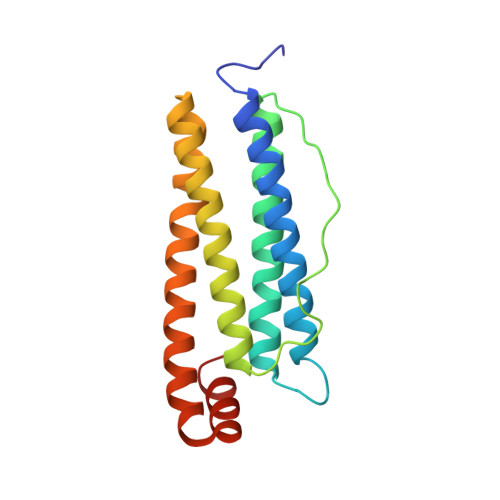
Mammalian ferritins are 24-mers assembled from two types of polypeptide chain which provide the molecule with different functions. H(eavy) chains catalyse the first step in iron storage, the oxidation of iron(II). L(ight) chains promote the nucleation of the mineral ferrihydrite enabling storage of iron(III) inside the protein shell. We report here the comparison of the three-dimensional structures of recombinant human H chain (HuHF) and horse L chain (HoLF) ferritin homopolymers, which have been refined at 1.9 A resolution. There is 53% sequence identity between these molecules, and the two structures are very similar, the H and L subunit alpha-carbons superposing to within 0.5 A rms deviation with 41 water molecules in common. Nevertheless, there are significant important differences which can be related to differences in function. In particular, the centres of the four-helix bundles contain distinctive groups of hydrophilic residues which have been associated with ferroxidase activity in H chains and enhanced stability in L chains. L chains contain a group of glutamates associated with mineralisation within the iron storage cavity of the protein.
- Krebs Institute for Biomolecular Research, Department of Molecular Biology and Biotechnology, University of Sheffield, England.
Organizational Affiliation: