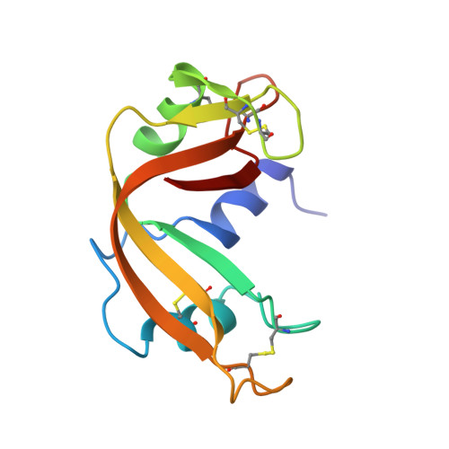
Crystal structures of ribonuclease A complexes with 5'-diphosphoadenosine 3'-phosphate and 5'-diphosphoadenosine 2'-phosphate at 1.7 A resolution.
Leonidas, D.D., Shapiro, R., Irons, L.I., Russo, N., Acharya, K.R.(1997) Biochemistry 36: 5578-5588
- PubMed: 9154942
- DOI: https://doi.org/10.1021/bi9700330
- Primary Citation of Related Structures:
1AFK, 1AFL, 1AFU - PubMed Abstract:
High-resolution (1.7 A) crystal structures have been determined for bovine pancreatic ribonuclease A (RNase A) complexed with 5'-diphosphoadenosine 3'-phosphate (ppA-3'-p) and 5'-diphosphoadenosine 2'-phosphate (ppA-2'-p), as well as for a native structure refined to 2.0 A. These nucleotide phosphates are the two most potent inhibitors of RNase A reported so far, with Ki values of 240 and 520 nM, respectively. The binding modes and conformations of ppA-3'-p and ppA-2'-p were found to differ markedly from those anticipated on the basis of earlier structures of RNase A complexes. The key difference is that the 5'-beta-phosphate rather than the 5'-alpha-phosphate of each inhibitor occupies the P1 phosphate binding site. As a consequence, the ribose moieties of the two nucleotides are shifted by approximately 2 A compared to the positions of their counterparts in earlier complexes, and the adenine rings are rotated into unusual syn conformations. Thus, the six-membered and five-membered rings of both adenines are reversed with respect to the others but nonetheless engage in extensive interactions with the residues that form the B2 purine binding site of RNase A. Despite the close structural similarity of the two inhibitors, the puckers of their furanose rings are different: C2'-endo and C3'-endo, respectively. Moreover, their 5'-alpha-phosphates and 3'(2')-monophosphates interact with largely different sets of RNase residues. The results of this crystallographic study emphasize the difficulties inherent in qualitative modeling of protein-inhibitor interactions and the compelling reasons for high-resolution structural studies in which quantitative design of improved inhibitors was enabled. The structures presented here provide a promising starting point for the rational design of tight-binding RNase inhibitors, which may be used as therapeutic agents in restraining the ribonucleolytic activities of RNase homologues such as angiogenin, eosinophil-derived neurotoxin, and eosinophil cationic protein.
- School of Biology and Biochemistry, University of Bath, Claverton Down, U.K.
Organizational Affiliation:


