1AJJ

LDL RECEPTOR LIGAND-BINDING MODULE 5, CALCIUM-COORDINATING


Experimental Data Snapshot

  • Method: X-RAY DIFFRACTION
  • Resolution: 1.70 Å
  • R-Value Free: 0.240 
  • R-Value Work: 0.209 
  • R-Value Observed: 0.209 

wwPDB Validation   3D Report Full Report


This is version 1.2 of the entry. See complete history


Literature

Molecular basis of familial hypercholesterolaemia from structure of LDL receptor module.

Fass, D.Blacklow, S.Kim, P.S.Berger, J.M.

(1997) Nature 388: 691-693

  • DOI: https://doi.org/10.1038/41798
  • Primary Citation of Related Structures:  
    1AJJ

  • PubMed Abstract: 

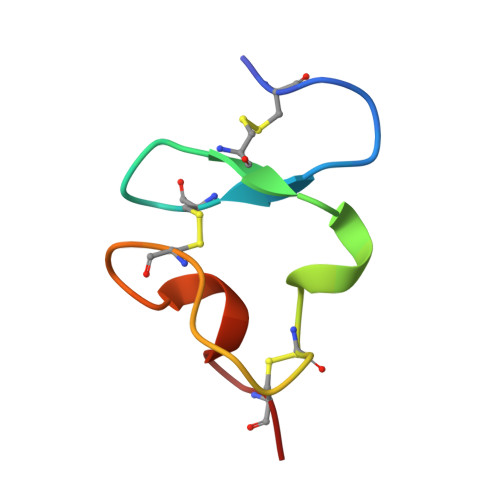
    The low-density lipoprotein receptor (LDLR) is responsible for the uptake of cholesterol-containing lipoprotein particles into cells. The amino-terminal region of LDLR, which consists of seven tandemly repeated, approximately 40-amino-acid, cysteine-rich modules (LDL-A modules), mediates binding to lipoproteins. LDL-A modules are biologically ubiquitous domains, found in over 100 proteins in the sequence database. The structure of ligand-binding repeat 5 (LR5) of the LDLR, determined to 1.7 A resolution by X-ray crystallography and presented here, contains a calcium ion coordinated by acidic residues that lie at the carboxy-terminal end of the domain and are conserved among LDL-A modules. Naturally occurring point mutations found in patients with the disease familial hypercholesterolaemia alter residues that directly coordinate Ca2+ or that serve as scaffolding residues of LR5.


  • Organizational Affiliation

    Howard Hughes Medical Institute, Department of Biology, Massachusetts Institute of Technology, Cambridge 02142, USA.


Macromolecules
Find similar proteins by:  (by identity cutoff)  |  3D Structure
Entity ID: 1
MoleculeChains Sequence LengthOrganismDetailsImage
LOW-DENSITY LIPOPROTEIN RECEPTOR37Homo sapiensMutation(s): 0 
UniProt & NIH Common Fund Data Resources
Find proteins for P01130 (Homo sapiens)
Explore P01130 
Go to UniProtKB:  P01130
PHAROS:  P01130
GTEx:  ENSG00000130164 
Entity Groups  
Sequence Clusters30% Identity50% Identity70% Identity90% Identity95% Identity100% Identity
UniProt GroupP01130
Sequence Annotations
Expand
  • Reference Sequence
Small Molecules
Ligands 2 Unique
IDChains Name / Formula / InChI Key2D Diagram3D Interactions
SO4
Query on SO4

Download Ideal Coordinates CCD File 
B [auth A]SULFATE ION
O4 S
QAOWNCQODCNURD-UHFFFAOYSA-L
CA
Query on CA

Download Ideal Coordinates CCD File 
C [auth A]CALCIUM ION
Ca
BHPQYMZQTOCNFJ-UHFFFAOYSA-N
Experimental Data & Validation

Experimental Data

  • Method: X-RAY DIFFRACTION
  • Resolution: 1.70 Å
  • R-Value Free: 0.240 
  • R-Value Work: 0.209 
  • R-Value Observed: 0.209 
  • Space Group: H 3
Unit Cell:
Length ( Å )Angle ( ˚ )
a = 53.45α = 90
b = 53.45β = 90
c = 26.76γ = 120
Software Package:
Software NamePurpose
DENZOdata reduction
SCALEPACKdata scaling
X-PLORmodel building
X-PLORrefinement
X-PLORphasing

Structure Validation

View Full Validation Report


Entry History 

Deposition Data

Revision History  (Full details and data files)

  • Version 1.0: 1997-07-07
    Type: Initial release
  • Version 1.1: 2008-03-24
    Changes: Version format compliance
  • Version 1.2: 2011-07-13
    Changes: Version format compliance