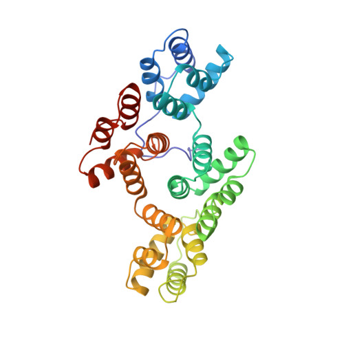
1AXN

THE HIGH RESOLUTION STRUCTURE OF ANNEXIN III SHOWS DIFFERENCES WITH ANNEXIN V


Experimental Data Snapshot

  • Method: X-RAY DIFFRACTION
  • Resolution: 1.78 Å
  • R-Value Free: 0.221 
  • R-Value Observed: 0.177 

wwPDB Validation   3D Report Full Report


This is version 1.3 of the entry. See complete history


Literature

The high-resolution crystal structure of human annexin III shows subtle differences with annexin V.

Favier-Perron, B.Lewit-Bentley, A.Russo-Marie, F.

(1996) Biochemistry 35: 1740-1744

  • DOI: https://doi.org/10.1021/bi952092o
  • Primary Citation of Related Structures:  
    1AXN

  • PubMed Abstract: 

    The structure of recombinant human annexin III was solved to 1.8 A resolution. Though homologous to annexin I and V, the annexin III structure shows significant differences. The tryptophan in the calcium loop of the third domain is exposed to the solvent, as in the structure of annexin V crystallized in high calcium concentrations, although the annexin III crystals were prepared at low calcium concentrations. The position of domain III relative to the other domains is different from both annexin V and I, suggesting further flexibility of the molecule. The entire N-terminus of the protein is well-defined in the present structure. The side chain of tryptophan 5 interacts with the hinge region of the hydrophillic channel, which could have an effect on the potential mobility of this region, as well as on its possible calcium channel behavior.


  • Organizational Affiliation

    ICGM, U332 INSERM, Paris, France.


Macromolecules
Find similar proteins by:  (by identity cutoff)  |  3D Structure
Entity ID: 1
MoleculeChains Sequence LengthOrganismDetailsImage
ANNEXIN III323Homo sapiensMutation(s): 0 
Gene Names: HUMAN ANNEXIN III
UniProt & NIH Common Fund Data Resources
Find proteins for P12429 (Homo sapiens)
Explore P12429 
Go to UniProtKB:  P12429
PHAROS:  P12429
GTEx:  ENSG00000138772 
Entity Groups  
Sequence Clusters30% Identity50% Identity70% Identity90% Identity95% Identity100% Identity
UniProt GroupP12429
Sequence Annotations
Expand
  • Reference Sequence
Experimental Data & Validation

Experimental Data

  • Method: X-RAY DIFFRACTION
  • Resolution: 1.78 Å
  • R-Value Free: 0.221 
  • R-Value Observed: 0.177 
  • Space Group: P 1 21 1
Unit Cell:
Length ( Å )Angle ( ˚ )
a = 42.544α = 90
b = 69.085β = 95.55
c = 50.95γ = 90
Software Package:
Software NamePurpose
PROLSQrefinement
MARXDSdata reduction

Structure Validation

View Full Validation Report


Entry History 

Deposition Data

Revision History  (Full details and data files)

  • Version 1.0: 1996-03-08
    Type: Initial release
  • Version 1.1: 2008-03-24
    Changes: Version format compliance
  • Version 1.2: 2011-07-13
    Changes: Version format compliance
  • Version 1.3: 2024-02-07
    Changes: Data collection, Database references, Derived calculations, Other