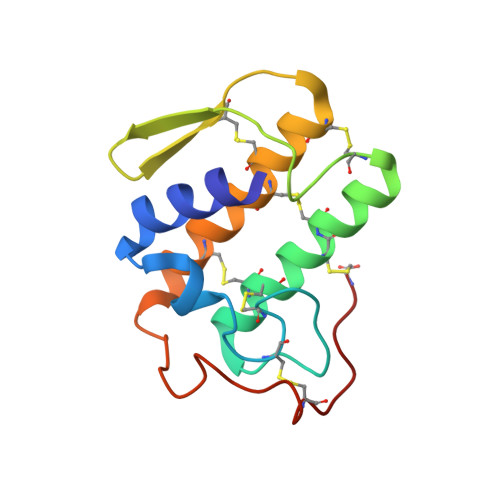
1AYP

A PROBE MOLECULE COMPOSED OF SEVENTEEN PERCENT OF TOTAL DIFFRACTING MATTER GIVES CORRECT SOLUTIONS IN MOLECULAR REPLACEMENT


Experimental Data Snapshot

  • Method: X-RAY DIFFRACTION
  • Resolution: 2.57 Å
  • R-Value Work: 0.214 
  • R-Value Observed: 0.214 

wwPDB Validation   3D Report Full Report


Ligand Structure Quality Assessment 

Created with Raphaël 2.3.0Worse 01 BetterLigand structure goodness of fit to experimental dataBest fitted INBClick on this verticalbar to view details

This is version 1.4 of the entry. See complete history


Literature

A probe molecule composed of seventeen percent of total diffracting matter gives correct solutions in molecular replacement.

Oh, B.H.

(1995) Acta Crystallogr D Biol Crystallogr 51: 140-144

  • DOI: https://doi.org/10.1107/S0907444994010024
  • Primary Citation of Related Structures:  
    1AYP

  • PubMed Abstract: 

    It is often found in the crystallization of enzyme-inhibitor complexes that an inhibitor causes crystal packing which is different to that of native protein. This is the case for crystals of human non-pancreatic secreted phospholipase A(2) (124 residues) containing six molecules in the asymmetric unit when the protein is complexed with a potential acylamino analogue of a phospholid. The hexameric structure was determined by molecular replacement using the structure of monomeric native protein as a probe. As an extension to the experiment, it was tested whether a backbone polypeptide composed of 17% of a known monomeric structure could find its correct position on a target molecule in molecular replacement. A probe model composed of the backbone atoms of the N-terminal 77 residues of lysine-, arginine-, ornithine-binding protein (LAO, a total of 238 residues) liganded with lysine correctly finds its position on LAO liganded with histidine which crystallizes as a monomer in the asymmetric unit. The results indicate that as little as 17% of total diffracting matter can be used in molecular replacement to solve crystal structures or to obtain phase information which can be combined with phases obtained by the isomorphous-replacement method.


  • Organizational Affiliation

    Department of Macromolecular Science, SmithKline Beecham Pharmaceuticals, King of Prussia, PA 19406, USA.


Macromolecules
Find similar proteins by:  (by identity cutoff)  |  3D Structure
Entity ID: 1
MoleculeChains Sequence LengthOrganismDetailsImage
PHOSPHOLIPASE A2
A, B, C, D, E
124Homo sapiensMutation(s): 0 
EC: 3.1.1.4
UniProt & NIH Common Fund Data Resources
Find proteins for P14555 (Homo sapiens)
Explore P14555 
Go to UniProtKB:  P14555
PHAROS:  P14555
GTEx:  ENSG00000188257 
Entity Groups  
Sequence Clusters30% Identity50% Identity70% Identity90% Identity95% Identity100% Identity
UniProt GroupP14555
Sequence Annotations
Expand
  • Reference Sequence
Small Molecules
Ligands 2 Unique
IDChains Name / Formula / InChI Key2D Diagram3D Interactions
INB
Query on INB

Download Ideal Coordinates CCD File 
I [auth A]
L [auth B]
O [auth C]
R [auth D]
U [auth E]
1-OCTADECYL-2-ACETAMIDO-2-DEOXY-SN-GLYCEROL-3-PHOSPHOETHYLMETHYL SULFIDE
C26 H54 N O6 P S
ZULTVWFLRZJENJ-AREMUKBSSA-N
CA
Query on CA

Download Ideal Coordinates CCD File 
G [auth A]
H [auth A]
J [auth B]
K [auth B]
M [auth C]
CALCIUM ION
Ca
BHPQYMZQTOCNFJ-UHFFFAOYSA-N
Experimental Data & Validation

Experimental Data

  • Method: X-RAY DIFFRACTION
  • Resolution: 2.57 Å
  • R-Value Work: 0.214 
  • R-Value Observed: 0.214 
  • Space Group: P 1 21 1
Unit Cell:
Length ( Å )Angle ( ˚ )
a = 64.51α = 90
b = 114.72β = 120.13
c = 64.43γ = 90
Software Package:
Software NamePurpose
X-PLORmodel building
X-PLORrefinement
X-PLORphasing

Structure Validation

View Full Validation Report


Ligand Structure Quality Assessment 

Created with Raphaël 2.3.0Worse 01 BetterLigand structure goodness of fit to experimental dataBest fitted INBClick on this verticalbar to view details

Entry History 

Deposition Data

  • Released Date: 1995-07-31 
  • Deposition Author(s): Oh, B.-H.

Revision History  (Full details and data files)

  • Version 1.0: 1995-07-31
    Type: Initial release
  • Version 1.1: 2008-03-24
    Changes: Version format compliance
  • Version 1.2: 2011-07-13
    Changes: Version format compliance
  • Version 1.3: 2024-06-05
    Changes: Data collection, Database references, Derived calculations, Other
  • Version 1.4: 2024-10-16
    Changes: Structure summary