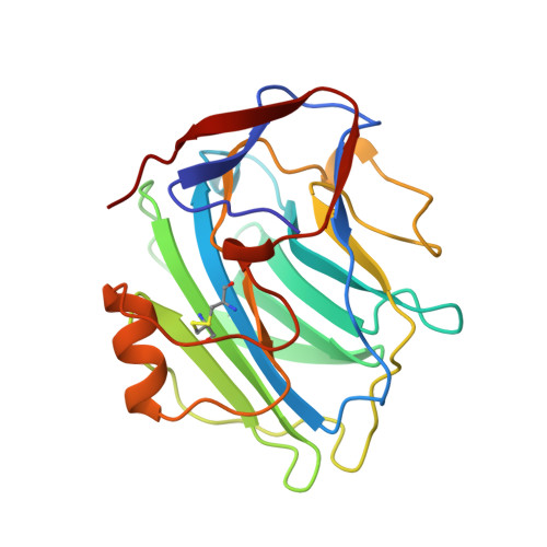
The physiological structure of human C-reactive protein and its complex with phosphocholine.
Thompson, D., Pepys, M.B., Wood, S.P.(1999) Structure 7: 169-177
- PubMed: 10368284
- DOI: https://doi.org/10.1016/S0969-2126(99)80023-9
- Primary Citation of Related Structures:
1B09 - PubMed Abstract:
Human C-reactive protein (CRP) is the classical acute phase reactant, the circulating concentration of which rises rapidly and extensively in a cytokine-mediated response to tissue injury, infection and inflammation. Serum CRP values are routinely measured, empirically, to detect and monitor many human diseases. However, CRP is likely to have important host defence, scavenging and metabolic functions through its capacity for calcium-dependent binding to exogenous and autologous molecules containing phosphocholine (PC) and then activating the classical complement pathway. CRP may also have pathogenic effects and the recent discovery of a prognostic association between increased CRP production and coronary atherothrombotic events is of particular interest. The X-ray structures of fully calcified C-reactive protein, in the presence and absence of bound PC, reveal that although the subunit beta-sheet jellyroll fold is very similar to that of the homologous pentameric protein serum amyloid P component, each subunit is tipped towards the fivefold axis. PC is bound in a shallow surface pocket on each subunit, interacting with the two protein-bound calcium ions via the phosphate group and with Glu81 via the choline moiety. There is also an unexpected hydrophobic pocket adjacent to the ligand. The structure shows how large ligands containing PC may be bound by CRP via a phosphate oxygen that projects away from the surface of the protein. Multipoint attachment of one planar face of the CRP molecule to a PC-bearing surface would leave available, on the opposite exposed face, the recognition sites for C1q, which have been identified by mutagenesis. This would enable CRP to target physiologically and/or pathologically significant complement activation. The hydrophobic pocket adjacent to bound PC invites the design of inhibitors of CRP binding that may have therapeutic relevance to the possible role of CRP in atherothrombotic events.
- School of Biological Sciences, University of Southampton, Southampton SO16 7PX UK.
Organizational Affiliation:


