1B0G | pdb_00001b0g

CLASS I HISTOCOMPATIBILITY ANTIGEN (HLA-A2.1)/BETA 2-MICROGLOBULIN/PEPTIDE P1049 COMPLEX


Experimental Data Snapshot

  • Method: X-RAY DIFFRACTION
  • Resolution: 2.50 Å
  • R-Value Free: 
    0.304 (Depositor), 0.288 (DCC) 
  • R-Value Work: 
    0.255 (Depositor), 0.246 (DCC) 
  • R-Value Observed: 
    0.260 (Depositor) 

Starting Model: experimental
View more details

wwPDB Validation 3D Report Full Report

Validation slider image for 1B0G

This is version 1.5 of the entry. See complete history

Literature

Structural evidence of T cell xeno-reactivity in the absence of molecular mimicry.

Zhao, R.Loftus, D.J.Appella, E.Collins, E.J.

(1999) J Exp Medicine 189: 359-370

  • DOI: https://doi.org/10.1084/jem.189.2.359
  • Primary Citation Related Structures: 
    1B0G, 1BZ9

  • PubMed Abstract: 

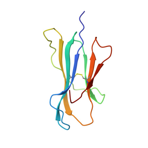
    The T cell receptor (TCR), from a xeno-reactive murine cytotoxic T lymphocyte clone AHIII12.2, recognizes murine H-2Db complexed with peptide p1027 (FAPGVFPYM), as well as human HLA-A2.1 complexed with peptide p1049 (ALWGFFPVL). A commonly proposed model (the molecular mimicry model) used to explain TCR cross-reactivity suggests that the molecular surfaces of the recognized complexes are similar in shape, charge, or both, in spite of the primary sequence differences. To examine the mechanism of xeno-reactivity of AHIII12.2, we have determined the crystal structures of A2/p1049 and Db/p1027 to 2.5 A and 2.8 A resolution, respectively. The crystal structures show that the TCR footprint regions of the two class I complexes are significantly different in shape and charge. We propose that rather than simple molecular mimicry, unpredictable arrays of common and differential contacts on the two class I complexes are used for their recognition by the same TCR.


  • Organizational Affiliation
    • Department of Microbiology and Immunology, University of North Carolina at Chapel Hill, Chapel Hill, North Carolina 27599, USA.

Macromolecule Content 

  • Total Structure Weight: 89.57 kDa 
  • Atom Count: 6,370 
  • Modeled Residue Count: 768 
  • Deposited Residue Count: 768 
  • Unique protein chains: 3

Macromolecules

Find similar proteins by:|  3D Structure
Entity ID: 1
MoleculeChains  Sequence LengthOrganismDetailsImage
CLASS I HISTOCOMPATIBILITY ANTIGEN
A, D
275Homo sapiensMutation(s): 0 
UniProt & NIH Common Fund Data Resources
Find proteins for P04439 (Homo sapiens)
Explore P04439 
Go to UniProtKB:  P04439
PHAROS:  P04439
GTEx:  ENSG00000206503 
Entity Groups
Sequence Clusters30% Identity50% Identity70% Identity90% Identity95% Identity100% Identity
UniProt GroupP04439
Sequence Annotations
Expand
Reference Sequence
Find similar proteins by:|  3D Structure
Entity ID: 2
MoleculeChains  Sequence LengthOrganismDetailsImage
BETA 2-MICROGLOBULIN
B, E
100Homo sapiensMutation(s): 0 
UniProt & NIH Common Fund Data Resources
Find proteins for P61769 (Homo sapiens)
Explore P61769 
Go to UniProtKB:  P61769
PHAROS:  P61769
GTEx:  ENSG00000166710 
Entity Groups
Sequence Clusters30% Identity50% Identity70% Identity90% Identity95% Identity100% Identity
UniProt GroupP61769
Sequence Annotations
Expand
Reference Sequence
Find similar proteins by:  Sequence   |   3D Structure  
Entity ID: 3
MoleculeChains  Sequence LengthOrganismDetailsImage
PEPTIDE P1049 (ALWGFFPVL)
C, F
9Homo sapiensMutation(s): 0 
Gene Names: EMC7
UniProt & NIH Common Fund Data Resources
Find proteins for Q9NPA0 (Homo sapiens)
Explore Q9NPA0 
Go to UniProtKB:  Q9NPA0
PHAROS:  Q9NPA0
GTEx:  ENSG00000134153 
Entity Groups
UniProt GroupQ9NPA0
Sequence Annotations
Expand
Reference Sequence

Experimental Data & Validation

Experimental Data

  • Method: X-RAY DIFFRACTION
  • Resolution: 2.50 Å
  • R-Value Free:  0.304 (Depositor), 0.288 (DCC) 
  • R-Value Work:  0.255 (Depositor), 0.246 (DCC) 
  • R-Value Observed: 0.260 (Depositor) 
Space Group: P 1
Unit Cell:
Length ( Å )Angle ( ˚ )
a = 50.09α = 81.98
b = 62.89β = 76.18
c = 74.68γ = 77.86
Software Package:
Software NamePurpose
DENZOdata reduction
SCALEPACKdata scaling
AMoREphasing
REFMACrefinement

Structure Validation

View Full Validation Report


Entry History 

Deposition Data

Revision History  (Full details and data files)

  • Version 1.0: 1998-11-18
    Type: Initial release
  • Version 1.1: 2008-04-26
    Changes: Version format compliance
  • Version 1.2: 2011-07-13
    Changes: Version format compliance
  • Version 1.3: 2013-07-10
    Changes: Other
  • Version 1.4: 2023-08-09
    Changes: Data collection, Database references, Refinement description
  • Version 1.5: 2024-11-13
    Changes: Structure summary