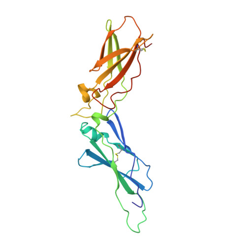
1BOY

EXTRACELLULAR REGION OF HUMAN TISSUE FACTOR


Experimental Data Snapshot

  • Method: X-RAY DIFFRACTION
  • Resolution: 2.20 Å
  • R-Value Work: 0.203 
  • R-Value Observed: 0.203 

wwPDB Validation   3D Report Full Report


This is version 1.3 of the entry. See complete history


Literature

Crystal structure of the extracellular region of human tissue factor.

Harlos, K.Martin, D.M.O'Brien, D.P.Jones, E.Y.Stuart, D.I.Polikarpov, I.Miller, A.Tuddenham, E.G.Boys, C.W.

(1994) Nature 370: 662-666

  • DOI: https://doi.org/10.1038/370662a0
  • Primary Citation of Related Structures:  
    1BOY

  • PubMed Abstract: 

    Tissue factor is a cell-surface glycoprotein receptor which initiates the blood coagulation cascade after vessel injury by interacting with blood clotting factor VII/VIIa and which is implicated in various pathological processes. When bound to tissue factor, factor VII is readily converted to the active protease factor VIIa by trace amounts of factors Xa, IXa or VIIa. Human tissue factor consists of 263 residues, the first 219 of which comprise the extracellular region. We have determined the crystal structure of the extracellular region at a resolution of 2.2 A. Tissue factor consists of two immunoglobulin-like domains associated through an extensive, novel, interdomain interface region. The binding site for factor VII lies at the interface region and involves residues from domain 1 and an extended loop (binding 'finger') of domain 2. This is the first reported structure of a representative of the class 2 cytokine receptor family, which also includes interferon-alpha, interferon-gamma (refs 2, 3) and interleukin-10 (ref. 4) receptors.


  • Organizational Affiliation

    Laboratory of Molecular Biophysics, Oxford Centre for Molecular Sciences, UK.


Macromolecules
Find similar proteins by:  (by identity cutoff)  |  3D Structure
Entity ID: 1
MoleculeChains Sequence LengthOrganismDetailsImage
HUMAN TISSUE FACTOR219Homo sapiensMutation(s): 0 
Gene Names: HUMAN TISSUE FACTOR CDNA CODON
UniProt & NIH Common Fund Data Resources
Find proteins for P13726 (Homo sapiens)
Explore P13726 
Go to UniProtKB:  P13726
PHAROS:  P13726
GTEx:  ENSG00000117525 
Entity Groups  
Sequence Clusters30% Identity50% Identity70% Identity90% Identity95% Identity100% Identity
UniProt GroupP13726
Sequence Annotations
Expand
  • Reference Sequence
Experimental Data & Validation

Experimental Data

  • Method: X-RAY DIFFRACTION
  • Resolution: 2.20 Å
  • R-Value Work: 0.203 
  • R-Value Observed: 0.203 
  • Space Group: P 41 21 2
Unit Cell:
Length ( Å )Angle ( ˚ )
a = 45.4α = 90
b = 45.4β = 90
c = 230.8γ = 90
Software Package:
Software NamePurpose
DENZOdata reduction
X-PLORrefinement
SCALEPACKdata scaling

Structure Validation

View Full Validation Report


Entry History 

Deposition Data

Revision History  (Full details and data files)

  • Version 1.0: 1996-06-10
    Type: Initial release
  • Version 1.1: 2008-03-03
    Changes: Version format compliance
  • Version 1.2: 2011-07-13
    Changes: Version format compliance
  • Version 1.3: 2024-10-30
    Changes: Data collection, Database references, Structure summary