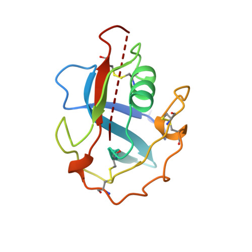
1BY2

STRUCTURE OF M2BP SCAVENGER RECEPTOR CYSTEINE-RICH DOMAIN


Experimental Data Snapshot

  • Method: X-RAY DIFFRACTION
  • Resolution: 2.00 Å
  • R-Value Free: 0.218 
  • R-Value Work: 0.186 
  • R-Value Observed: 0.186 

wwPDB Validation   3D Report Full Report


This is version 1.4 of the entry. See complete history


Literature

Crystal structure of a scavenger receptor cysteine-rich domain sheds light on an ancient superfamily.

Hohenester, E.Sasaki, T.Timpl, R.

(1999) Nat Struct Biol 6: 228-232

  • DOI: https://doi.org/10.1038/6669
  • Primary Citation of Related Structures:  
    1BY2

  • PubMed Abstract: 

    Scavenger receptor cysteine-rich (SRCR) domains are found widely in cell surface molecules and in some secreted proteins, where they are thought to mediate ligand binding. We have determined the crystal structure at 2.0 A resolution of the SRCR domain of Mac-2 binding protein (M2BP), a tumor-associated antigen and matrix protein. The structure reveals a curved six-stranded beta-sheet cradling an alpha-helix. Structure-based sequence alignment demonstrates that the M2BP SRCR domain is a valid template for the entire SRCR protein superfamily. This allows an interpretation of previous mutagenesis data on ligand binding to the lymphocyte receptor CD6.


  • Organizational Affiliation

    Department of Crystallography, Birkbeck College, University of London, UK.


Macromolecules
Find similar proteins by:  (by identity cutoff)  |  3D Structure
Entity ID: 1
MoleculeChains Sequence LengthOrganismDetailsImage
MAC-2 BINDING PROTEIN119Homo sapiensMutation(s): 0 
UniProt & NIH Common Fund Data Resources
Find proteins for Q08380 (Homo sapiens)
Explore Q08380 
Go to UniProtKB:  Q08380
PHAROS:  Q08380
GTEx:  ENSG00000108679 
Entity Groups  
Sequence Clusters30% Identity50% Identity70% Identity90% Identity95% Identity100% Identity
UniProt GroupQ08380
Glycosylation
Glycosylation Sites: 1Go to GlyGen: Q08380-1
Sequence Annotations
Expand
  • Reference Sequence
Small Molecules
Ligands 1 Unique
IDChains Name / Formula / InChI Key2D Diagram3D Interactions
NAG
Query on NAG

Download Ideal Coordinates CCD File 
B [auth A]2-acetamido-2-deoxy-beta-D-glucopyranose
C8 H15 N O6
OVRNDRQMDRJTHS-FMDGEEDCSA-N
Experimental Data & Validation

Experimental Data

  • Method: X-RAY DIFFRACTION
  • Resolution: 2.00 Å
  • R-Value Free: 0.218 
  • R-Value Work: 0.186 
  • R-Value Observed: 0.186 
  • Space Group: P 43 21 2
Unit Cell:
Length ( Å )Angle ( ˚ )
a = 44.63α = 90
b = 44.63β = 90
c = 134.54γ = 90
Software Package:
Software NamePurpose
MOSFLMdata reduction
SCALAdata scaling
CCP4model building
X-PLORrefinement
CCP4data scaling
CCP4phasing

Structure Validation

View Full Validation Report


Entry History 

Deposition Data

Revision History  (Full details and data files)

  • Version 1.0: 1999-05-18
    Type: Initial release
  • Version 1.1: 2008-03-24
    Changes: Version format compliance
  • Version 1.2: 2011-07-13
    Changes: Non-polymer description, Version format compliance
  • Version 1.3: 2020-07-29
    Type: Remediation
    Reason: Carbohydrate remediation
    Changes: Advisory, Data collection, Derived calculations, Structure summary
  • Version 1.4: 2024-11-20
    Changes: Advisory, Data collection, Database references, Structure summary