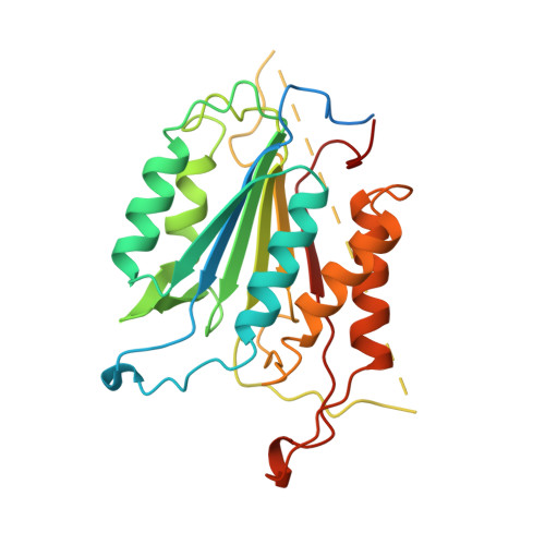
1CP3

CRYSTAL STRUCTURE OF THE COMPLEX OF APOPAIN WITH THE TETRAPEPTIDE INHIBITOR ACE-DVAD-FMC


Experimental Data Snapshot

  • Method: X-RAY DIFFRACTION
  • Resolution: 2.30 Å
  • R-Value Free: 0.284 
  • R-Value Work: 0.188 
  • R-Value Observed: 0.188 

Starting Model: in silico
View more details

wwPDB Validation   3D Report Full Report


This is version 1.5 of the entry. See complete history


Literature

Structure of recombinant human CPP32 in complex with the tetrapeptide acetyl-Asp-Val-Ala-Asp fluoromethyl ketone.

Mittl, P.R.Di Marco, S.Krebs, J.F.Bai, X.Karanewsky, D.S.Priestle, J.P.Tomaselli, K.J.Grutter, M.G.

(1997) J Biol Chem 272: 6539-6547

  • DOI: https://doi.org/10.1074/jbc.272.10.6539
  • Primary Citation of Related Structures:  
    1CP3

  • PubMed Abstract: 

    The cysteine protease CPP32 has been expressed in a soluble form in Escherichia coli and purified to >95% purity. The three-dimensional structure of human CPP32 in complex with the irreversible tetrapeptide inhibitor acetyl-Asp-Val-Ala-Asp fluoromethyl ketone was determined by x-ray crystallography at a resolution of 2.3 A. The asymmetric unit contains a (p17/p12)2 tetramer, in agreement with the tetrameric structure of the protein in solution as determined by dynamic light scattering and size exclusion chromatography. The overall topology of CPP32 is very similar to that of interleukin-1beta-converting enzyme (ICE); however, differences exist at the N terminus of the p17 subunit, where the first helix found in ICE is missing in CPP32. A deletion/insertion pattern is responsible for the striking differences observed in the loops around the active site. In addition, the P1 carbonyl of the ketone inhibitor is pointing into the oxyanion hole and forms a hydrogen bond with the peptidic nitrogen of Gly-122, resulting in a different state compared with the tetrahedral intermediate observed in the structure of ICE and CPP32 in complex with an aldehyde inhibitor. The topology of the interface formed by the two p17/p12 heterodimers of CPP32 is different from that of ICE. This results in different orientations of CPP32 heterodimers compared with ICE heterodimers, which could affect substrate recognition. This structural information will be invaluable for the design of small synthetic inhibitors of CPP32 as well as for the design of CPP32 mutants.


  • Organizational Affiliation

    Core Drug Discovery Technologies, Ciba-Geigy AG, CH-4002 Basel, Switzerland.


Macromolecules
Find similar proteins by:  (by identity cutoff)  |  3D Structure
Entity ID: 1
MoleculeChains Sequence LengthOrganismDetailsImage
APOPAINA,
C [auth B]
277Homo sapiensMutation(s): 0 
EC: 3.4.22 (PDB Primary Data), 3.4.22.56 (UniProt)
UniProt & NIH Common Fund Data Resources
Find proteins for P42574 (Homo sapiens)
Explore P42574 
Go to UniProtKB:  P42574
PHAROS:  P42574
GTEx:  ENSG00000164305 
Entity Groups  
Sequence Clusters30% Identity50% Identity70% Identity90% Identity95% Identity100% Identity
UniProt GroupP42574
Sequence Annotations
Expand
  • Reference Sequence

Find similar proteins by:  Sequence   |   3D Structure  

Entity ID: 2
MoleculeChains Sequence LengthOrganismDetailsImage
ACETYL-ASP-VAL-ALA-ASP-FLUOROMETHYLKETONEB [auth C],
D
6N/AMutation(s): 0 
Sequence Annotations
Expand
  • Reference Sequence
Experimental Data & Validation

Experimental Data

  • Method: X-RAY DIFFRACTION
  • Resolution: 2.30 Å
  • R-Value Free: 0.284 
  • R-Value Work: 0.188 
  • R-Value Observed: 0.188 
  • Space Group: P 1 21 1
Unit Cell:
Length ( Å )Angle ( ˚ )
a = 50.9α = 90
b = 69.1β = 101.2
c = 93.8γ = 90
Software Package:
Software NamePurpose
AMoREphasing
X-PLORrefinement
MARXDSdata reduction
MARSCALEdata scaling

Structure Validation

View Full Validation Report


Entry History 

Deposition Data

Revision History  (Full details and data files)

  • Version 1.0: 1997-12-24
    Type: Initial release
  • Version 1.1: 2008-03-24
    Changes: Version format compliance
  • Version 1.2: 2011-07-13
    Changes: Atomic model, Database references, Derived calculations, Non-polymer description, Structure summary, Version format compliance
  • Version 1.3: 2012-12-12
    Changes: Other
  • Version 1.4: 2024-04-03
    Changes: Data collection, Database references, Derived calculations, Refinement description
  • Version 1.5: 2024-10-09
    Changes: Structure summary