1D0G

CRYSTAL STRUCTURE OF DEATH RECEPTOR 5 (DR5) BOUND TO APO2L/TRAIL


Experimental Data Snapshot

  • Method: X-RAY DIFFRACTION
  • Resolution: 2.40 Å
  • R-Value Free: 0.267 
  • R-Value Work: 0.222 

wwPDB Validation   3D Report Full Report


This is version 1.3 of the entry. See complete history


Literature

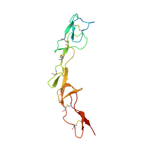
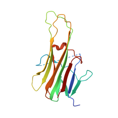
Triggering cell death: the crystal structure of Apo2L/TRAIL in a complex with death receptor 5.

Hymowitz, S.G.Christinger, H.W.Fuh, G.Ultsch, M.O'Connell, M.Kelley, R.F.Ashkenazi, A.de Vos, A.M.

(1999) Mol Cell 4: 563-571

  • DOI: https://doi.org/10.1016/s1097-2765(00)80207-5
  • Primary Citation of Related Structures:  
    1D0G

  • PubMed Abstract: 

    Formation of a complex between Apo2L (also called TRAIL) and its signaling receptors, DR4 and DR5, triggers apoptosis by inducing the oligomerization of intracellular death domains. We report the crystal structure of the complex between Apo2L and the ectodomain of DR5. The structure shows three elongated receptors snuggled into long crevices between pairs of monomers of the homotrimeric ligand. The interface is divided into two distinct patches, one near the bottom of the complex close to the receptor cell surface and one near the top. Both patches contain residues that are critical for high-affinity binding. A comparison to the structure of the lymphotoxin-receptor complex suggests general principles of binding and specificity for ligand recognition in the TNF receptor superfamily.


  • Organizational Affiliation

    Department of Protein Engineering, Genentech, Inc., South San Francisco, California 94080, USA.


Macromolecules
Find similar proteins by:  (by identity cutoff)  |  3D Structure
Entity ID: 1
MoleculeChains Sequence LengthOrganismDetailsImage
DEATH RECEPTOR-5A [auth R],
B [auth S],
C [auth T]
130Homo sapiensMutation(s): 0 
UniProt & NIH Common Fund Data Resources
Find proteins for O14763 (Homo sapiens)
Explore O14763 
Go to UniProtKB:  O14763
PHAROS:  O14763
GTEx:  ENSG00000120889 
Entity Groups  
Sequence Clusters30% Identity50% Identity70% Identity90% Identity95% Identity100% Identity
UniProt GroupO14763
Sequence Annotations
Expand
  • Reference Sequence
Find similar proteins by:  (by identity cutoff)  |  3D Structure
Entity ID: 2
MoleculeChains Sequence LengthOrganismDetailsImage
APOPTOSIS-2 LIGANDD [auth A],
E [auth B],
F [auth D]
168Homo sapiensMutation(s): 0 
UniProt & NIH Common Fund Data Resources
Find proteins for P50591 (Homo sapiens)
Explore P50591 
Go to UniProtKB:  P50591
PHAROS:  P50591
GTEx:  ENSG00000121858 
Entity Groups  
Sequence Clusters30% Identity50% Identity70% Identity90% Identity95% Identity100% Identity
UniProt GroupP50591
Sequence Annotations
Expand
  • Reference Sequence
Experimental Data & Validation

Experimental Data

  • Method: X-RAY DIFFRACTION
  • Resolution: 2.40 Å
  • R-Value Free: 0.267 
  • R-Value Work: 0.222 
  • Space Group: P 21 21 21
Unit Cell:
Length ( Å )Angle ( ˚ )
a = 66.818α = 90
b = 111.019β = 90
c = 130.807γ = 90
Software Package:
Software NamePurpose
DENZOdata reduction
SCALEPACKdata scaling
AMoREphasing
X-PLORrefinement

Structure Validation

View Full Validation Report


Entry History 

Deposition Data

Revision History  (Full details and data files)

  • Version 1.0: 1999-10-22
    Type: Initial release
  • Version 1.1: 2008-04-27
    Changes: Version format compliance
  • Version 1.2: 2011-07-13
    Changes: Version format compliance
  • Version 1.3: 2018-01-31
    Changes: Advisory, Experimental preparation