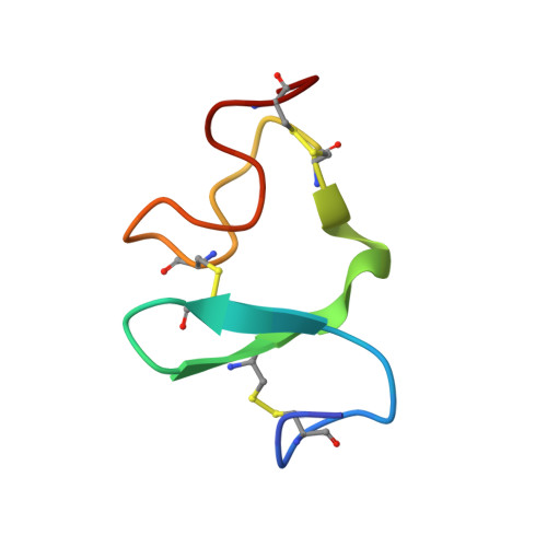
Solution structure of the sixth LDL-A module of the LDL receptor.
North, C.L., Blacklow, S.C.(2000) Biochemistry 39: 2564-2571
- PubMed: 10704205 Search on PubMed
- DOI: https://doi.org/10.1021/bi992087a
- Primary Citation Related Structures:
1D2J - PubMed Abstract:
The low-density lipoprotein receptor (LDLR) is the primary mechanism for uptake of plasma cholesterol into cells and serves as a prototype for an entire class of cell surface receptors. The amino-terminal domain of the receptor consists of seven LDL-A modules; the third through the seventh modules all contribute to the binding of low-density lipoproteins (LDLs). Here, we present the NMR solution structure of the sixth LDL-A module (LR6) from the ligand binding domain of the LDLR. This module, which has little recognizable secondary structure, retains the essential structural features observed in the crystal structure of LDL-A module five (LR5) of the LDLR. Three disulfide bonds, a pair of buried residues forming a hydrophobic "mini-core", and a calcium-binding site that serves to organize the C-terminal lobe of the module all occupy positions in LR6 similar to those observed in LR5. The striking presence of a conserved patch of negative surface electrostatic potential among LDL-A modules of known structure suggests that ligand recognition by these repeats is likely to be mediated in part by electrostatic complementarity of receptor and ligand. Two variants of LR6, identified originally as familial hypercholesterolemia (FH) mutations, have been investigated for their ability to form native disulfide bonds under conditions that permit disulfide exchange. The first, E219K, lies near the amino-terminal end of LR6, whereas the second, D245E, alters one of the aspartate side chains that directly coordinate the bound calcium ion. After equilibration at physiologic calcium concentrations, neither E219K nor D245E folds to a unique disulfide isomer, indicating that FH mutations both within and distant from the calcium-binding site give rise to protein-folding defects.
- Department of Pathology, Brigham and Women's Hospital and Harvard Medical School, Boston, Massachusetts 02115, USA.
Organizational Affiliation: