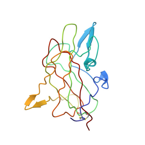
1D7P

Crystal structure of the c2 domain of human factor viii at 1.5 a resolution at 1.5 A


Experimental Data Snapshot

  • Method: X-RAY DIFFRACTION
  • Resolution: 1.50 Å
  • R-Value Free: 0.215 
  • R-Value Work: 0.205 
  • R-Value Observed: 0.206 

wwPDB Validation   3D Report Full Report


This is version 1.5 of the entry. See complete history


Literature

Structure of the C2 domain of human factor VIII at 1.5 A resolution.

Pratt, K.P.Shen, B.W.Takeshima, K.Davie, E.W.Fujikawa, K.Stoddard, B.L.

(1999) Nature 402: 439-442

  • DOI: https://doi.org/10.1038/46601
  • Primary Citation of Related Structures:  
    1D7P

  • PubMed Abstract: 

    Human factor VIII is a plasma glycoprotein that has a critical role in blood coagulation. Factor VIII circulates as a complex with von Willebrand factor. After cleavage by thrombin, factor VIIIa associates with factor IXa at the surface of activated platelets or endothelial cells. This complex activates factor X (refs 6, 7), which in turn converts prothrombin to thrombin in the presence of factor Va (refs 8, 9). The carboxyl-terminal C2 domain of factor VIII contains sites that are essential for its binding to von Willebrand factor and to negatively charged phospholipid surfaces. Here we report the structure of human factor VIII C2 domain at 1.5 A resolution. The structure reveals a beta-sandwich core, from which two beta-turns and a loop display a group of solvent-exposed hydrophobic residues. Behind the hydrophobic surface lies a ring of positively charged residues. This motif suggests a mechanism for membrane binding involving both hydrophobic and electrostatic interactions. The structure explains, in part, mutations in the C2 region of factor VIII that lead to bleeding disorders in haemophilia A.


  • Organizational Affiliation

    Program in Structural Biology, Division of Basic Sciences, Fred Hutchinson Cancer Research Center, Seattle, Washington 98109, USA.


Macromolecules
Find similar proteins by:  (by identity cutoff)  |  3D Structure
Entity ID: 1
MoleculeChains Sequence LengthOrganismDetailsImage
COAGULATION FACTOR VIII PRECURSORA [auth M]159Homo sapiensMutation(s): 1 
Membrane Entity: Yes 
UniProt & NIH Common Fund Data Resources
Find proteins for P00451 (Homo sapiens)
Explore P00451 
Go to UniProtKB:  P00451
PHAROS:  P00451
GTEx:  ENSG00000185010 
Entity Groups  
Sequence Clusters30% Identity50% Identity70% Identity90% Identity95% Identity100% Identity
UniProt GroupP00451
Sequence Annotations
Expand
  • Reference Sequence
Experimental Data & Validation

Experimental Data

  • Method: X-RAY DIFFRACTION
  • Resolution: 1.50 Å
  • R-Value Free: 0.215 
  • R-Value Work: 0.205 
  • R-Value Observed: 0.206 
  • Space Group: P 21 21 21
Unit Cell:
Length ( Å )Angle ( ˚ )
a = 46.44α = 90
b = 56.41β = 90
c = 65.74γ = 90
Software Package:
Software NamePurpose
DENZOdata reduction
SCALEPACKdata scaling
SHARPphasing
CNSrefinement

Structure Validation

View Full Validation Report


Entry History 

Deposition Data

Revision History  (Full details and data files)

  • Version 1.0: 1999-12-01
    Type: Initial release
  • Version 1.1: 2008-04-27
    Changes: Version format compliance
  • Version 1.2: 2011-07-13
    Changes: Non-polymer description, Version format compliance
  • Version 1.3: 2021-11-03
    Changes: Database references, Derived calculations
  • Version 1.4: 2023-12-13
    Changes: Data collection, Derived calculations
  • Version 1.5: 2024-10-30
    Changes: Structure summary