1DQB | pdb_00001dqb

NMR STRUCTURE OF THROMBOMODULIN EGF(4-5)


Experimental Data Snapshot

  • Method: SOLUTION NMR
  • Conformers Calculated: 50 
  • Conformers Submitted: 12 
  • Selection Criteria: structures with the least restraint violations 

wwPDB Validation 3D Report Full Report

Validation slider image for 1DQB

This is version 1.4 of the entry. See complete history

Literature

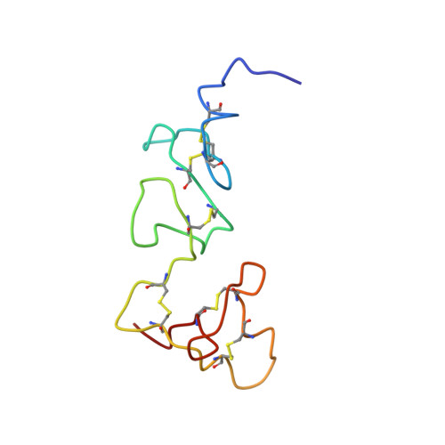
Solution structure of the smallest cofactor-active fragment of thrombomodulin.

Wood, M.J.Sampoli-Benitez, B.A.Komives, E.A.

(2000) Nat Struct Biol 7: 200-204

  • DOI: https://doi.org/10.1038/73302
  • Primary Citation Related Structures: 
    1DQB

  • PubMed Abstract: 

    A glycosylated fragment of thrombomodulin containing two epidermal growth factor-like domains (TMEGF45) was analyzed by NMR. The 4th-domains structure of this two-domain fragment is similar to that of the individual domain previously determined. The 5th-domain, which has uncrossed disulfide bonds, is not as well determined in the two-domain fragment than the individual domain previously solved. The flexibility of the 5th-domain is consistent with low heteronuclear NOEs. In the individual 5th-domain, Met 388 was disordered, and key thrombin binding residues formed a hydrophobic core. By contrast, in TMEGF45, Met 388 is in the 5th-domain core, positioned by Phe 376 from the 4th-domain. As a result, key thrombin binding residues that were in the core of the individual domain are expelled. Upon thrombin binding, chemical shifts of two residues in the 4th-domain, the three interdomain linker residues, and nearly all of the 5th-domain are perturbed. Thus, TMEGF45 binds thrombin by an induced fit mechanism involving a flexible 5th-domain.


  • Organizational Affiliation
    • Department of Chemistry and Biochemistry, University of California, San Diego, 9500 Gilman Dr., La Jolla, California 92093-0359, USA.

Macromolecule Content 

  • Total Structure Weight: 9.74 kDa 
  • Atom Count: 669 
  • Modeled Residue Count: 83 
  • Deposited Residue Count: 83 
  • Unique protein chains: 1

Macromolecules

Find similar proteins by:|  3D Structure
Entity ID: 1
MoleculeChains  Sequence LengthOrganismDetailsImage
THROMBOMODULIN83Homo sapiensMutation(s): 0 
UniProt & NIH Common Fund Data Resources
Find proteins for P07204 (Homo sapiens)
Explore P07204 
Go to UniProtKB:  P07204
PHAROS:  P07204
GTEx:  ENSG00000178726 
Entity Groups
Sequence Clusters30% Identity50% Identity70% Identity90% Identity95% Identity100% Identity
UniProt GroupP07204
Glycosylation
Glycosylation Sites: 2Go to GlyGen: P07204-1
Sequence Annotations
Expand
Reference Sequence

Small Molecules

Ligands 1 Unique
IDChains Name / Formula / InChI Key2D Diagram3D Interactions
NAG

Query on NAG


Download:Ideal Coordinates CCD File
B [auth A],
C [auth A]
2-acetamido-2-deoxy-beta-D-glucopyranose
C8 H15 N O6
OVRNDRQMDRJTHS-FMDGEEDCSA-N

Experimental Data & Validation

Experimental Data

  • Method: SOLUTION NMR
  • Conformers Calculated: 50 
  • Conformers Submitted: 12 
  • Selection Criteria: structures with the least restraint violations 

Structure Validation

View Full Validation Report


Entry History 

Deposition Data

Revision History  (Full details and data files)

  • Version 1.0: 2000-03-06
    Type: Initial release
  • Version 1.1: 2008-04-27
    Changes: Version format compliance
  • Version 1.2: 2011-07-13
    Changes: Non-polymer description, Version format compliance
  • Version 1.3: 2020-07-29
    Type: Remediation
    Reason: Carbohydrate remediation
    Changes: Data collection, Database references, Derived calculations, Structure summary
  • Version 1.4: 2024-10-16
    Changes: Data collection, Database references, Structure summary