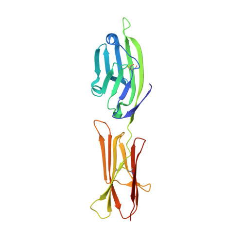
1DR9

CRYSTAL STRUCTURE OF A SOLUBLE FORM OF B7-1 (CD80)


Experimental Data Snapshot

  • Method: X-RAY DIFFRACTION
  • Resolution: 3.00 Å
  • R-Value Free: 0.280 
  • R-Value Work: 0.238 
  • R-Value Observed: 0.238 

wwPDB Validation   3D Report Full Report


This is version 1.6 of the entry. See complete history


Literature

Structure and dimerization of a soluble form of B7-1.

Ikemizu, S.Gilbert, R.J.Fennelly, J.A.Collins, A.V.Harlos, K.Jones, E.Y.Stuart, D.I.Davis, S.J.

(2000) Immunity 12: 51-60

  • DOI: https://doi.org/10.1016/s1074-7613(00)80158-2
  • Primary Citation of Related Structures:  
    1DR9

  • PubMed Abstract: 

    B7-1 (CD80) and B7-2 (CD86) are glycoproteins expressed on antigen-presenting cells. The binding of these molecules to the T cell homodimers CD28 and CTLA-4 (CD152) generates costimulatory and inhibitory signals in T cells, respectively. The crystal structure of the extracellular region of B7-1 (sB7-1), solved to 3 A resolution, consists of a novel combination of two Ig-like domains, one characteristic of adhesion molecules and the other previously seen only in antigen receptors. In the crystal lattice, sB7-1 unexpectedly forms parallel, 2-fold rotationally symmetric homodimers. Analytical ultracentrifugation reveals that sB7-1 also dimerizes in solution. The structural data suggest a mechanism whereby the avidity-enhanced binding of B7-1 and CTLA-4 homodimers, along with the relatively high affinity of these interactions, favors the formation of very stable inhibitory signaling complexes.


  • Organizational Affiliation

    Division of Structural Biology, Wellcome Trust Centre for Human Genetics, The University of Oxford, United Kingdom.


Macromolecules
Find similar proteins by:  (by identity cutoff)  |  3D Structure
Entity ID: 1
MoleculeChains Sequence LengthOrganismDetailsImage
T LYMPHOCYTE ACTIVATION ANTIGEN201Homo sapiensMutation(s): 1 
Gene Names: CHO
UniProt & NIH Common Fund Data Resources
Find proteins for P33681 (Homo sapiens)
Explore P33681 
Go to UniProtKB:  P33681
PHAROS:  P33681
GTEx:  ENSG00000121594 
Entity Groups  
Sequence Clusters30% Identity50% Identity70% Identity90% Identity95% Identity100% Identity
UniProt GroupP33681
Glycosylation
Glycosylation Sites: 3Go to GlyGen: P33681-1
Sequence Annotations
Expand
  • Reference Sequence
Experimental Data & Validation

Experimental Data

  • Method: X-RAY DIFFRACTION
  • Resolution: 3.00 Å
  • R-Value Free: 0.280 
  • R-Value Work: 0.238 
  • R-Value Observed: 0.238 
  • Space Group: I 41 2 2
Unit Cell:
Length ( Å )Angle ( ˚ )
a = 57.28α = 90
b = 57.28β = 90
c = 298.93γ = 90
Software Package:
Software NamePurpose
DENZOdata reduction
SCALEPACKdata scaling
SOLVEphasing
CNSrefinement

Structure Validation

View Full Validation Report


Entry History 

Deposition Data

Revision History  (Full details and data files)

  • Version 1.0: 2000-01-10
    Type: Initial release
  • Version 1.1: 2008-04-27
    Changes: Version format compliance
  • Version 1.2: 2011-07-13
    Changes: Non-polymer description, Version format compliance
  • Version 1.3: 2018-01-31
    Changes: Experimental preparation
  • Version 1.4: 2020-07-29
    Type: Remediation
    Reason: Carbohydrate remediation
    Changes: Data collection, Derived calculations, Structure summary
  • Version 1.5: 2021-11-03
    Changes: Database references, Structure summary
  • Version 1.6: 2024-10-16
    Changes: Data collection, Structure summary