1DSU | pdb_00001dsu

HUMAN FACTOR D, COMPLEMENT ACTIVATING ENZYME


Experimental Data Snapshot

  • Method: X-RAY DIFFRACTION
  • Resolution: 2.00 Å
  • R-Value Free: 
    0.203 (Depositor) 
  • R-Value Work: 
    0.188 (Depositor), 0.196 (DCC) 
  • R-Value Observed: 
    0.188 (Depositor) 

wwPDB Validation 3D Report Full Report

Validation slider image for 1DSU

This is version 1.4 of the entry. See complete history

Literature

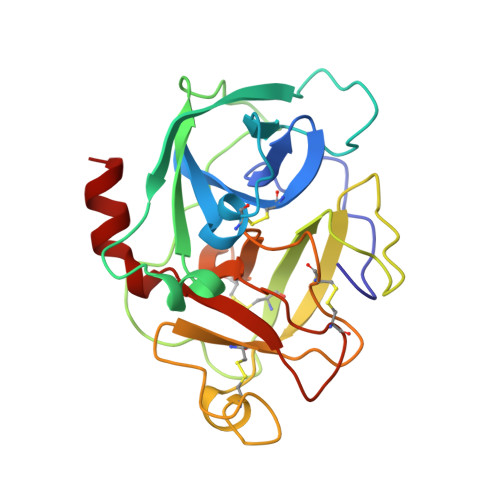
Structure of human factor D. A complement system protein at 2.0 A resolution.

Narayana, S.V.Carson, M.el-Kabbani, O.Kilpatrick, J.M.Moore, D.Chen, X.Bugg, C.E.Volanakis, J.E.DeLucas, L.J.

(1994) J Mol Biology 235: 695-708

  • DOI: https://doi.org/10.1006/jmbi.1994.1021
  • Primary Citation Related Structures: 
    1DSU

  • PubMed Abstract: 

    Factor D, an essential enzyme for the activation of the alternative pathway of the complement system, belongs to the serine protease superfamily. The crystal structure of the enzyme was solved by a combination of multiple isomorphous replacement and molecular replacement methods. The present model was refined to an R-factor of 18.8% using 23,681 observed reflections between 7.5 and 2.0 A resolution, with a root-mean-square deviation from standard bond lengths of 0.016 A. The two non-crystallographically related molecules in the triclinic unit cell have distinctive active site conformations. The protein has the general structural fold of a serine protease, but there are several unique amino acid substitutions resulting in significant alterations in the critical loops responsible for catalysis and substrate specificity in serine proteases. Factor D is the first complement serine protease whose three-dimensional structure has been determined.


  • Organizational Affiliation
    • Center for Macromolecular Crystallography, University of Alabama at Birmingham 35294.

Macromolecule Content 

  • Total Structure Weight: 48.88 kDa 
  • Atom Count: 3,499 
  • Modeled Residue Count: 456 
  • Deposited Residue Count: 456 
  • Unique protein chains: 1

Macromolecules

Find similar proteins by:|  3D Structure
Entity ID: 1
MoleculeChains  Sequence LengthOrganismDetailsImage
FACTOR D
A, B
228Homo sapiensMutation(s): 0 
EC: 3.4.21.46
UniProt & NIH Common Fund Data Resources
Find proteins for P00746 (Homo sapiens)
Explore P00746 
Go to UniProtKB:  P00746
PHAROS:  P00746
GTEx:  ENSG00000197766 
Entity Groups
Sequence Clusters30% Identity50% Identity70% Identity90% Identity95% Identity100% Identity
UniProt GroupP00746
Sequence Annotations
Expand
Reference Sequence

Experimental Data & Validation

Experimental Data

  • Method: X-RAY DIFFRACTION
  • Resolution: 2.00 Å
  • R-Value Free:  0.203 (Depositor) 
  • R-Value Work:  0.188 (Depositor), 0.196 (DCC) 
  • R-Value Observed: 0.188 (Depositor) 
Space Group: P 1
Unit Cell:
Length ( Å )Angle ( ˚ )
a = 40.99α = 101.26
b = 65.02β = 109.89
c = 40.36γ = 74.32
Software Package:
Software NamePurpose
XENGENdata collection
X-PLORmodel building
X-PLORrefinement
XENGENdata reduction
X-PLORphasing

Structure Validation

View Full Validation Report


Entry History 

Deposition Data

Revision History  (Full details and data files)

  • Version 1.0: 1996-08-17
    Type: Initial release
  • Version 1.1: 2008-03-25
    Changes: Version format compliance
  • Version 1.2: 2011-07-13
    Changes: Version format compliance
  • Version 1.3: 2024-06-05
    Changes: Data collection, Database references, Other
  • Version 1.4: 2024-10-23
    Changes: Structure summary