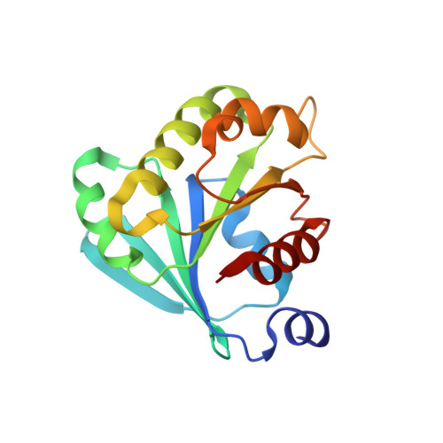
1E0S

small G protein Arf6-GDP


Experimental Data Snapshot

  • Method: X-RAY DIFFRACTION
  • Resolution: 2.28 Å
  • R-Value Free: 0.234 
  • R-Value Work: 0.172 
  • R-Value Observed: 0.172 

Starting Model: experimental
View more details

wwPDB Validation   3D Report Full Report


Ligand Structure Quality Assessment 


This is version 1.4 of the entry. See complete history


Literature

Structure of Arf6-Gdp Suggests a Basis for Guanine Nucleotide Exchange Factors Specificity

Menetrey, J.Macia, E.Pasqualato, S.Franco, M.Cherfils, J.

(2000) Nat Struct Biol 7: 466

  • DOI: https://doi.org/10.1038/75863
  • Primary Citation of Related Structures:  
    1E0S

  • PubMed Abstract: 

    Arf6 is an isoform of Arf that localizes at the periphery of the cell where it has an essential role in endocytotic pathways. Its function does not overlap with that of Arf1, although the two proteins share approximately 70% sequence identity and they have switch regions, whose conformation depends on the nature of the guanine nucleotide, with almost identical sequences. The crystal structure of Arf6-GDP at 2.3 A shows that it has a conformation similar to that of Arf1-GDP, which cannot bind membranes with high affinity. Significantly, the switch regions of Arf6 deviate by 2-5 A from those of Arf1. These differences are a consequence of the shorter N-terminal linker of Arf6 and of discrete sequence changes between Arf6 and Arf1. Mutational analysis shows that one of the positions which differs between Arf1 and Arf6 affects the configuration of the nucleotide binding site and thus the nucleotide binding properties of the Arf variant. Altogether, our results provide a structural basis for understanding how Arf1 and Arf6 can be distinguished by their guanine nucleotide exchange factors and suggest a model for the nucleotide/membrane cycle of Arf6.


  • Organizational Affiliation

    Laboratoire d'Enzymologie et Biochimie Structurales, CNRS, Gif-sur-Yvette, France.


Macromolecules
Find similar proteins by:  (by identity cutoff)  |  3D Structure
Entity ID: 1
MoleculeChains Sequence LengthOrganismDetailsImage
ADP-ribosylation factor 6174Homo sapiensMutation(s): 0 
Gene Names: ARF6
EC: 3.6.5.2
UniProt & NIH Common Fund Data Resources
Find proteins for P62330 (Homo sapiens)
Explore P62330 
Go to UniProtKB:  P62330
PHAROS:  P62330
GTEx:  ENSG00000165527 
Entity Groups  
Sequence Clusters30% Identity50% Identity70% Identity90% Identity95% Identity100% Identity
UniProt GroupP62330
Sequence Annotations
Expand
  • Reference Sequence
Experimental Data & Validation

Experimental Data

  • Method: X-RAY DIFFRACTION
  • Resolution: 2.28 Å
  • R-Value Free: 0.234 
  • R-Value Work: 0.172 
  • R-Value Observed: 0.172 
  • Space Group: P 61 2 2
Unit Cell:
Length ( Å )Angle ( ˚ )
a = 55.66α = 90
b = 55.66β = 90
c = 194.95γ = 120
Software Package:
Software NamePurpose
X-PLORrefinement
DENZOdata reduction
SCALEPACKdata scaling
AMoREphasing

Structure Validation

View Full Validation Report


Ligand Structure Quality Assessment 


Entry History 

Deposition Data

Revision History  (Full details and data files)

  • Version 1.0: 2000-04-18
    Type: Initial release
  • Version 1.1: 2011-05-08
    Changes: Version format compliance
  • Version 1.2: 2011-07-13
    Changes: Version format compliance
  • Version 1.3: 2019-09-25
    Changes: Data collection, Database references, Derived calculations, Other, Source and taxonomy, Structure summary
  • Version 1.4: 2023-12-06
    Changes: Data collection, Database references, Derived calculations, Refinement description